
Cellular Imaging Systems
Video Gallery


Featured videos and webinars
3D Imaging Seminar Series
Plate Annotations in CRX
ImageXpress Pico : Acquisition and Analysis Basics
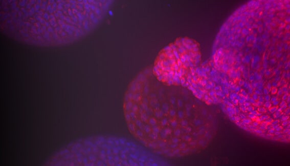
Intestinal organoids for automated screening assays
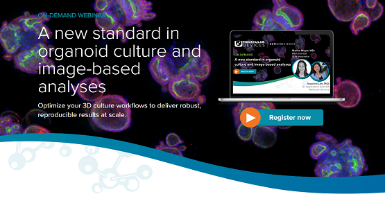
New standard in organoid culture and image-based analyses
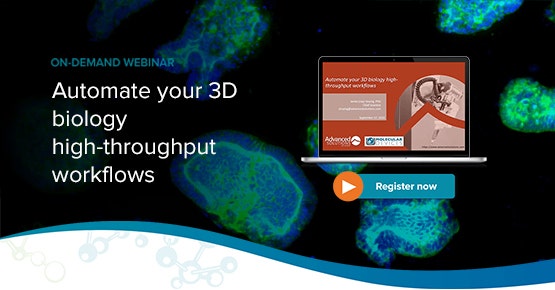
Automate your 3D biology high-throughput workflows

How 3D biology is shaping the future of drug discovery
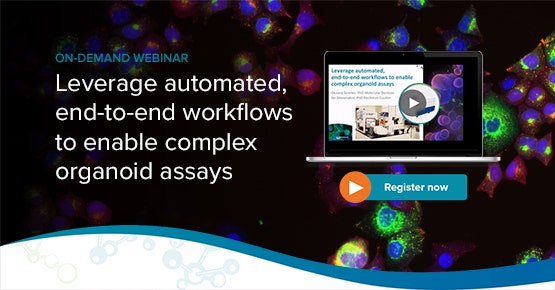
Leverage automated, end-to-end workflows to enable complex organoid assays
Automation of the organ-on-a-chip assay: automated culture, imaging and analysis of angiogenesis
Challenges for drug screening of complex biological systems
3D Tissue Models Imaging and Automation of Organ-On-A-Chip
Introduction to the Organoid Innovation Center and ImageXpress Confocal HT.ai system
Level Up your 3D Cell Culture: From Research to High-Throughput
Enhance high-content 3D biology imaging with automated sample preparation
Enhancing 3D Disease Models: Automated, High-Throughput, Phenotypic Screening with Organ-on-a-Chip
High-Content Phenotypic Screening
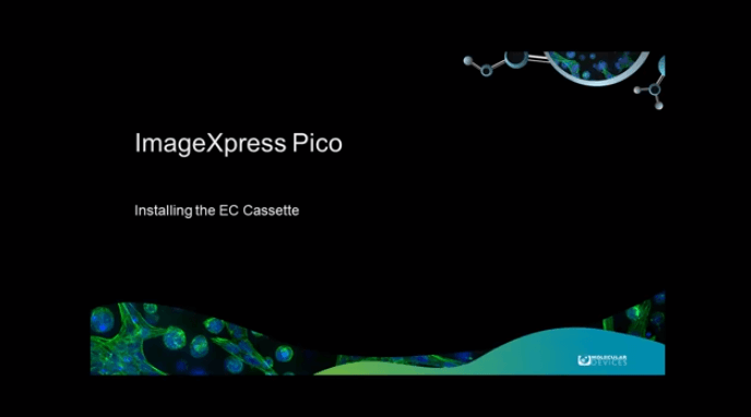
ImageXpress Pico - Installing the EC Cassette
3D Imaging Seminar Series: Fast and simple 3D spheroid analysis using automated high-content imaging
Emerging Organoid Models: Translating Basic Research to Drug Development and Regenerative Medicine
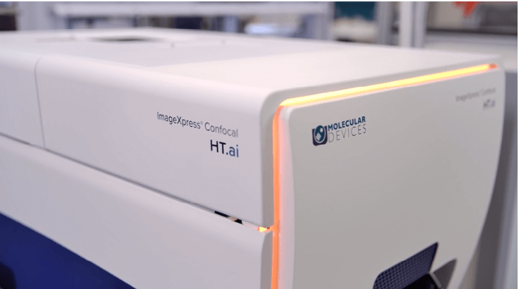
ImageXpress Confocal HT.ai Overview
ImageXpress Confocal HT.ai Overview
https://share.vidyard.com/watch/B4KG58gz5Ar4bc4snph4go
ImageXpress Confocal HT.ai / IN Carta Image Analysis Software Introduction
The ImageXpress® Confocal HT.ai High-Content Imaging System utilizes a seven-channel laser light source with eight imaging channels to enable highly multiplexed assays while maintaining high throughput by using shortened exposure times.
https://share.vidyard.com/watch/QHSgGc52W62U73JC9Muiz2
Getting Started with 3D Human Tissue Models and Imaging
https://share.vidyard.com/watch/A1WircbtdV7wgR1Pqr39yu
Capturing the Complexity of Cell Biology
https://share.vidyard.com/watch/WrM2vds8jA6Ayc2joqUDnR
Organoid Innovation Center Walkthrough
ImageXpress Micro Confocal High-Content Imaging System
https://share.vidyard.com/watch/b3RtIKRdaR7Nc6VowrGtNg
ImageXpress Micro Confocal System
The ImageXpress® Micro Confocal system is a high-content solution that can switch between widefield and confocal imaging of fixed and live cells.

An AI-based approach to high-content phenotypic characterization of human iPSC-derviced neuronal cells
https://share.vidyard.com/watch/24FeCTPpMYqMJUCLauSFTD
ImageXpress Micro Confocal Virtual Tour

Disease modeling in the 21st century: Automated organoid assays with 3D imaging

3D cell culture, tissue clearing, & high-content imaging in the quest of effective solutions to NAFLD
ImageXpress Micro 4 High-Content Imaging System
https://share.vidyard.com/watch/0KdmdV--9VL3H3OYY7miyw
ImageXpress Micro 4 System
The ImageXpress® Micro 4 High-Content Imaging System is a high-throughput, widefield imager that can acquire images of whole organisms and cellular or intracellular events.
https://share.vidyard.com/watch/UI3mt4vhsfG8ELC8Z3QJBw
Pushing the Boundaries of High Content Screening
https://share.vidyard.com/watch/5Hz2HkvJ47JnHRaWzl0q0w
Preparing Assays for Genome-wide RNAi Screens Using High Content Microscopy
https://share.vidyard.com/watch/hk_BRvQClBcPPNh5twAXAA
Multiplexed High Content Hepatoxicity Assays Using iPSC-Derived Hepatocytes
https://share.vidyard.com/watch/diDVJ30m_-JgqiYPNf-MIw
High Content Imaging Analysis of Cell Sheet Morphogenesis Utilizing in Vitro Tissue Models
ImageXpress Nano Automated Imaging System
https://share.vidyard.com/watch/AEGF6og2cXN8HPphQNYhqJ
ImageXpress Nano High-Content Imaging System Virtual Tour
Virtual tour of the ImageXpress Nano, including hardware and software which demonstrates the image acquisition as well as image review and analysis.

Accelerate your screening with high-content and automated imaging

Accelerating study of viral infection and therapeutics with microplate-based detection and high-throughput screening
https://share.vidyard.com/watch/VQLtB1d26JmCYvi2VYJUtA
LabTube Meets Molecular Devices & MIMETAS, Susan Murphy & Sebastiaan Trietsch

Magnetic 3D Bioprinting, 3D Cell Culture in a 2D Workflow
ImageXpress Pico Automated Cell Imaging System
https://share.vidyard.com/watch/Z3iBwKY6jzq5BkFsqDfjBi
ImageXpress Pico System
The ImageXpress® Pico Automated Cell Imaging System is more than a digital microscope, combining high-resolution imaging with powerful analysis.
https://share.vidyard.com/watch/nJKT2dgwt3UJGDL5EZwkoD
ImageXpress Pico Automated Imaging System Virtual Tour

Automated Imaging and You – Quantitative microscopy for every lab, Powerful data for all

https://view.ceros.com/molecular-devices/imagexpress-pico-digital-confocal/p/1
ImageXpress Pico Automated Cell Imaging System – Feature Update
https://share.vidyard.com/watch/dVSCYeXmvjoqyR9UvhVCyM
A Product Manager's Tour of the ImageXpress Pico
https://share.vidyard.com/watch/Z3iBwKY6jzq5BkFsqDfjBi
ImageXpress Pico System
The ImageXpress® Pico Automated Cell Imaging System is more than a digital microscope, combining high-resolution imaging with powerful analysis.
https://share.vidyard.com/watch/nJKT2dgwt3UJGDL5EZwkoD
ImageXpress Pico Automated Imaging System Virtual Tour

Ein Einstieg in die Bildgebung von 3D-Zellmodellen – alles, was Sie wissen müssen
https://view.ceros.com/molecular-devices/imagexpress-pico-digital-confocal/p/1
ImageXpress Pico Automated Cell Imaging System – Feature Update
https://share.vidyard.com/watch/dVSCYeXmvjoqyR9UvhVCyM
A Product Manager's Tour of the ImageXpress Pico
https://share.vidyard.com/watch/Z3iBwKY6jzq5BkFsqDfjBi
ImageXpress Pico System
The ImageXpress® Pico Automated Cell Imaging System is more than a digital microscope, combining high-resolution imaging with powerful analysis.
https://share.vidyard.com/watch/nJKT2dgwt3UJGDL5EZwkoD
ImageXpress Pico Automated Imaging System Virtual Tour

L'imagerie automatisée et vous - La microscopie quantitative pour chaque laboratoire, un puissant outil pour l’analyse et la visualisation de données

Bien démarrer avec l'imagerie de modèles cellulaires 3D - tout ce que vous devez savoir
https://view.ceros.com/molecular-devices/imagexpress-pico-digital-confocal/p/1
ImageXpress Pico Automated Cell Imaging System – Feature Update
https://share.vidyard.com/watch/dVSCYeXmvjoqyR9UvhVCyM
A Product Manager's Tour of the ImageXpress Pico
CellReporterXpress Image Acquisition and Analysis Software
https://share.vidyard.com/watch/jrvgKE4G3VTLhZcCqZwqnb
How to perform a z-stack image acquisition using CellReporterXpress software
The CellReporterXpress Automated Image Acquisition and Analysis Software works with the ImageXpress® Pico system. It has a simple, easy-to-learn interface for performing quantitative analysis on images acquired from automated microscopy.
https://share.vidyard.com/watch/KPtbYfqE5nuduV2eJiKuTL
How to easily image slides and regions of interest with Live Preview on the ImageXpress Pico system
https://share.vidyard.com/watch/iwwbwYap13jMxP8XuEVrDG
Configuring environmental control settings on the ImageXpress Pico system
https://share.vidyard.com/watch/e7fDVbVU9qaETHjwZaahji
Transmitted light cell scoring on the ImageXpress Pico
https://share.vidyard.com/watch/QU7cyvFrkykG4MYPYmNGPk
Setting up acquisition and analysis on the ImageXpress Pico
MetaXpress High-Content Image Acquisition and Analysis Software
https://share.vidyard.com/watch/vWpmKGGNMGDMUkVh3Fd35b
Basic workflow from image acquisition to analysis on the ImageXpress Micro Confocal system using MetaXpress software
MetaXpress® High-Content Image Acquisition and Analysis Software is a comprehensive solution for high-content analysis featuring a tightly orchestrated and integrated workflow.
https://share.vidyard.com/watch/eU6diFWP4io5Hyo4uoJdBm
Plate annotation and curve fitting in MetaXpress software
https://share.vidyard.com/watch/G3SsKxYAmNbCpoLjPNwHRd
Quickstart Guide: Plate acquisition on the ImageXpress Micro Confocal system using MetaXpress software
https://share.vidyard.com/watch/39eTiBo59htbFbzFSYau9h
Implement High-Throughput 3D Image Analysis for Samples from Subcellular Structures to Spheroids
https://share.vidyard.com/watch/Bs1SecHnFPiHyJ6Lp5S8Qs
Quickstart Guide: Review images on the ImageXpress Micro Confocal system using MetaXpress software
MetaMorph Microscopy Automation and Image Analysis Software
https://share.vidyard.com/watch/OQGNWMI_cUjQX3k1PuKHDQ
Resolving Molecular Organization and Dynamics Using Localization-Based Super-Resolution Microscopy
MetaMorph® Microscopy Automation and Image Analysis Software automates acquisition, device control, and image analysis. It easily integrates dissimilar fluorescent microscope hardware and peripherals into a single custom workstation.
https://share.vidyard.com/watch/DCmR2tZopOHTpRu5aNPbRQ
Real-Time Single-Molecule Based Super-Resolution Microscopy Reconstruction: Theoretical and Practical Insight