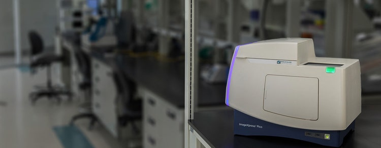

ImageXpress Pico Automated Cell Imaging System
몇 분 만에 샘플에서 결과 확인까지 진행할 수 있는 세포 이미징 및 분석 시스템
자동 Brightfield, 형광 및 Digital Confocal 이미징의 디지털 현미경
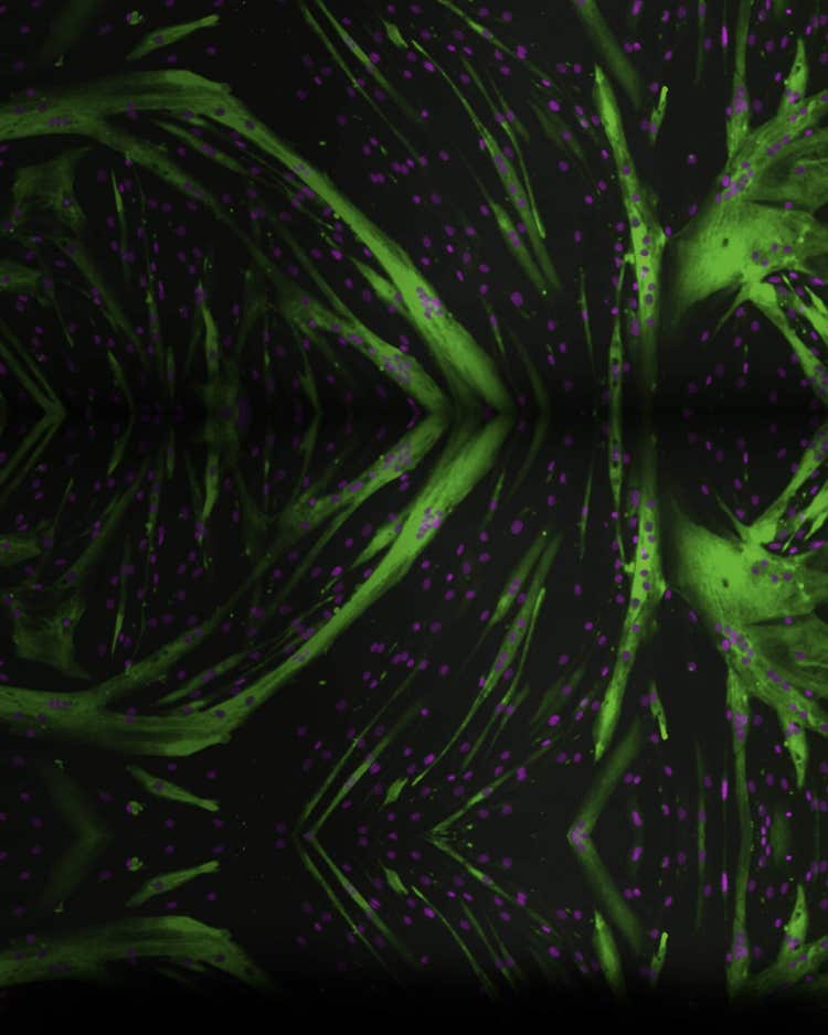
ImageXpress® Pico Automated Cell Imaging System은 고해상도 이미징을 강력한 분석과 결합하여 디지털 현미경보다 우수합니다. 형광 이미징 또는 Brightfield assay를 실행하는 경우, 자동화 이미저에 포함된 종합적이며 사전 구성된 Cell Based Assay 프로토콜 포트폴리오가 학습 곡선을 단축하므로 실험 실행을 빠르게 시작할 수 있습니다. Digital Confocal* 2D On-The-Fly 데콘볼루션, 자동 초점, Live Preview, 다중 파장 세포 점수화, 선택형 IN Carta® 이미지 분석 소프트웨어 실험과정과 같은 기능을 갖춘 ImageXpress Pico는 소형이며 합리적인 가격의 이미저로 발견의 수준을 높일 수 있도록 지원해 드립니다.

신속하게 시작
아이콘 중점, 사용자 친화적인 CellReporterXpress® 이미지 획득 및 분석 소프트웨어를 사용하면 전체 실험실이 디지털 현미경 관찰을 간소화할 수 있습니다. 최소한의 교육만으로 이미지 포착 및 분석을 시작할 수 있습니다.

Cell Counting 이상의 효과 획득
세포사멸, 미토콘드리아 평가, 3d Cell Model, Live Cell/Timelapse, 다중 파장 Cell Scoring 및 Neurite tracing 등 다양한 세포 기반 실험에 최적화된 25개 이상의 사전 구성 템플릿으로 assay를 확장할 수 있습니다.

합리적인 비용의 이미징 자동화
샘플을 실행하기 위해 핵심 실험실에 가야 하는 번거로움에서 해방될 수 있습니다. 이 시스템의 실험실 친화적인 가격을 통해, 연구원들은 실험실 벤치에서 자동화 이미징과 분석의 편의성을 누릴 수 있습니다. Digital Confocal, environmental control 및 z-stack 획득과 같은 옵션을 통해, 시스템을 연구에 적합하게 주문할 수 있습니다.
기능

다양한 이미징 모드
ImageXpress Pico 시스템은 4x에서 63x의 대물렌즈를 제공하며, Colorimetirc, Brightfield, 형광 또는 Digital Confocal 2D On-the-fly Deconvolution 이미징 모드로 작동할 수 있습니다.

내장된 이미지 분석 프로토콜
단순한 Cell Counting부터 정교한 Neurite Tracing 분석에 이르기까지 다양한 25개 이상의 내장 분석 프로토콜이 있습니다. 클릭 기능 검색 도구와 같은 기능을 통해, 특정 기준에 맞는 몇 개의 세포를 클릭하기만 해도 분석 Parameter를 최적화할 수 있습니다.

Plate-to-individual Cell 보기
플레이트 전체보기에서 개별 세포보기까지 여러 수준에서 데이타를 시각화할 수 있습니다. 이러한 다양한 데이터 시각화 도구는 사용자가 다양한 레벨의 이미지와 assay로부터 최대한 많은 정보를 얻을 수 있도록 도와줍니다.

Z-Stack 이미지 획득
z-stack 획득을 사용하여 좀 더 정확한 세분화를 위한 더욱 선명한 이미지를 생성할 수 있습니다. 서로 다른 초점에서 일련의 이미지를 획득하여 단일 절편(Slice)보다 좀 더 세부적인 정보를 얻을 수 있습니다. 사용자들은 모든 절편을 포함시키거나 최종 이미지에 어떤 절편을 포함시킬 것인지 선택할 수 있습니다.

관심 영역(ROI)을 신속하고 손쉽게 식별
Live Preview 기능을 사용하면 사용자가 샘플을 가상 조이스틱을 이용하여 초점을 조정할 수 있으므로 관심영역(ROI)을 확인하는 과정이 간편해지고, 빠르고 손쉽게 작업할 수 있습니다.

EC(Environmental Control)
장기간 동안 수행되는 Time Lapse 및 Live Cell Assay는 습도, CO2 및 O2 제어를 위한 옵션이 포함된 Environmental Control System을 사용하여 실행할 수 있습니다. 또한 z 이동을 방지하기 위해 최적화된 이 소프트웨어는 환경 상태를 실시간으로 모니터링하여, 최적의 assay 조건을 보장합니다.
ImageXpress Pico와 CellReporterXpress의 강력한 결합 경험
최신 자료
제품 응용 분야
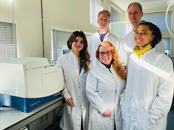
고객 사례 소개