모든 실험실에서 접근할 수 있는 형광 이미징 플랫폼
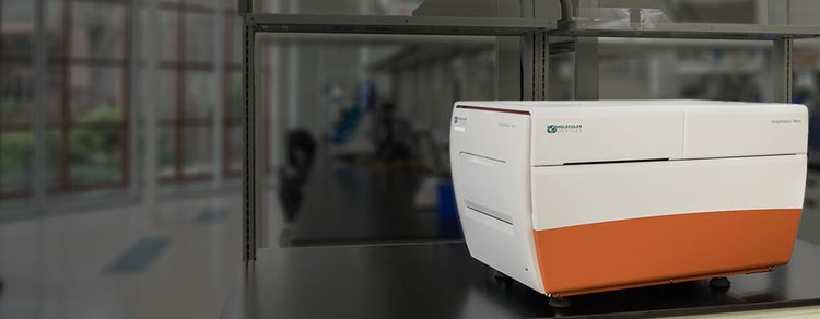
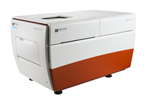
ImageXpress® Nano 자동화 Imaging 시스템은 적절한 assay 감도를 안정적으로 제공하기 위한 수명이 긴 고체 상태의 조명 엔진 및 광학 장치를 특징으로 합니다. 이 강력하고 유연한 형광 현미경 관찰 솔루션으로 다양한 세포 및 세포하 assay에 대한 미세한 세부 정보를 포착할 수 있습니다. 이 시스템에는 2D 및 3D Imaging 및 Timelapse 분석과 사용 편의성부터 독점적인 assay 설계까지 다양한 요구 사항을 위한 도구를 갖춘 MetaXpress® High-Content Image 획득 및 분석 소프트웨어가 포함됩니다.

Label-Free 이미징
Brightfield 이미징으로 유해한 형광물질을 사용하지 않고도 빠른 획득이 가능합니다.

이미지 분석 간소화
MetaXpress® 소프트웨어의 모듈 도구함을 사용하면 수백 개의 일상적 assay를 신속하게 설정할 수 있습니다. 더 우수한 편의성을 위한 턴키 응용 분야 모듈로 구성된 당사의 옵션 선택 사항 중에서 선택하십시오.

다양한 샘플 포착
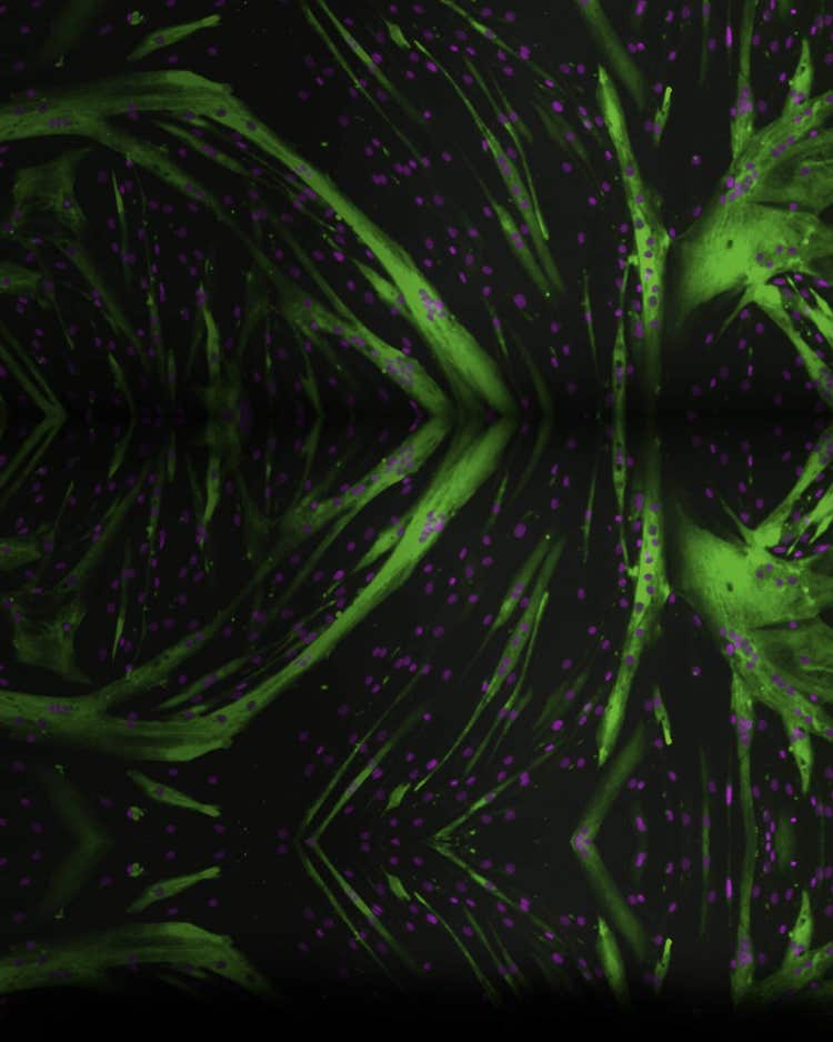
이 시스템은 2x에서 60x의 배율로 전체 Well(C. elegans, 제브라피시)과 세부 내 정보(vesicle, 세포소기관)를 이미징하기 위한 유연성을 제공합니다.
기능

넓은 시야
384 Well 플레이트의 전체 Well은 더 빠른 처리를 위해 4x 배율의 단일 이미지로 포착할 수 있습니다.

자동화된 스테이지
해상도가 25nm 이상인 완전 자동화된 X, Y 및 Z 스테이지입니다.

다양한 필터 및 대물렌즈
이 시스템은 연구 요구 사항에 맞도록 다양한 필터 또는 대물렌즈(2~60x)로 구성할 수 있습니다.

다섯 가지 형광 채널
이 시스템은 한 번에 최대 5개의 형광 필터를 설치할 수 있습니다. 이 소프트웨어는 한 번에 최대 7개의 채널을 획득할 수 있어 하나의 실험에서 다채널 형광과 Transmitted Light Imaging을 지원합니다.
고속 자동 초점
레이저 자동 초점을 통해 플레이트, 슬라이드 및 불규칙한 표면 전체에서 빠르고 일관된 초점을 맞출 수 있습니다.
EC(Environmental Control)
장기간 실험하는 Timelapse 및 Live cell assay는 온도, 습도 및 CO2 제어를 위한 옵션이 포함된 온 보드 환경 시스템을 사용하여 실행할 수 있습니다.
최신 자료
제품 응용 분야
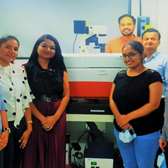
고객 사례 소개