

ImageXpress Micro Confocal High-Content Imaging 시스템
1주일에 백만 개 이상의 Well을 이미징하는 기능을 갖춘 Unique Confocal Imaging Solution
Water immersion 대물 옵션이 포함된 high-content confocal imaging 솔루션
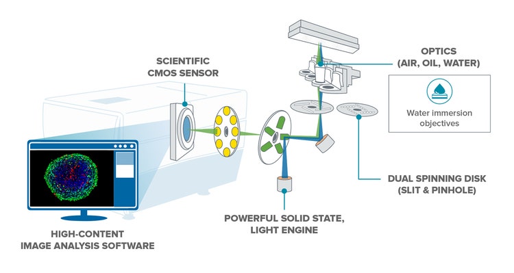
ImageXpress® Micro Confocal 시스템은 Fixed 및 Live Cell의 Widefield 및 Confocal Imaging 간에 전환할 수 있는 high-content 솔루션입니다. 전체 유기체, 두꺼운 조직, 2D 및 3D 모델, 그리고 세포 또는 세포 내 사건의 고품질 이미지를 포착할 수 있습니다. 스피닝 디스크 공초점 및 sCMOS 카메라는 심장세포 박동과 줄기세포 분화와 같이 빠른 속도로 드물게 발생하는 사건의 Imaging을 지원합니다. 이 시스템은 MetaXpress 소프트웨어와 Water Immersion Objective와 같은 유연한 옵션을 선택할 수 있어, 3D assay 개발에서 스크리닝에 이르기까지 다양한 confocal imaging 응용 분야를 지원합니다.

더 높은 품질의 이미지 획득
당사의 독점적인 AgileOptix™ 스피닝 디스크 공초점 기술, 광시야 및 밝은 광원으로 우수한 대비, high resolution 이미지를 포착할 수 있습니다.

이미지 획득 및 분석 맞춤화
획득과 분석 Parameter를 궁극적으로 제어할 수 있으므로, 3D 구조 분석에서 유기체 또는 세포의 Population 내에 있는 특정 대물의 표적 이미징에 이르기까지 다양한 응용 분야를 지원합니다. Water Immersion Objective, 세포 환경 제어 또는 당사의 고성능 맞춤화와 같은 표준 옵션으로 더 많은 응용 분야를 실행할 수 있습니다.

더 짧은 시간에 더 많은 데이터 분석
MetaXpress® PowerCore™ 소프트웨어는 High-throughput 환경에서 분석 속도를 가속화합니다. 이 소프트웨어는 다중 CPU 환경으로 이미지 처리 작업을 분산합니다.
기능

넓은 동적 범위(Dynamic Range)
>3의 로그 동적 범위 세기 검출을 통한 단일 이미지의 낮고 높은 세기 신호 정량화입니다.

독점적인 AgileOptix 스피닝 디스크 공초점
이 기술은 특수 설계 광학 장치, 고출력 고체 조명 엔진 및 sCMOS 센서로 향상된 감도를 제공합니다. 교체 가능 디스크 기하학적 구조는 속도와 해상도 사이에 유연성을 제공합니다.

광시야
광시야는 전체 Well confocal imaging을 지원하고 누락된 표적을 제거합니다.

옵션 온 보드 로봇 유체공학 장치
복합물 첨가, 웰 세척 및 배지 교환이 관련되는 assay의 경우, 옵션 온 보드 로봇 유체공학 장치를 사용할 수 있습니다.

정확한 3D 측정
MetaXpress 3D 분석 모듈은 confocal imaging에 맞게 최적화되어 있기 때문에 부피와 거리에 대한 3D 측정을 지원합니다.

다양한 이미징 모드
이 시스템은 Phase Contrast 및 Brightfield Label Free Imaging, Florescence, Widefield, Colorimetric, Confocal Imaging과 Immersion Imaging을 표준 옵션으로 제공합니다.
AgileOptix™ 기술
ImageXpress Micro 공초점 시스템은 당사의 독점 AgileOptix 기술을 통해 까다로운 응용 분야에 필요한 민감도와 처리량을 제공합니다. AgileOptix는 강력한 고체 상태 광원, 특별히 제작된 광학적 장치, 과학적 CMOS 센서, 디스크의 기하학적 구조를 변화시키는 능력을 결합합니다.
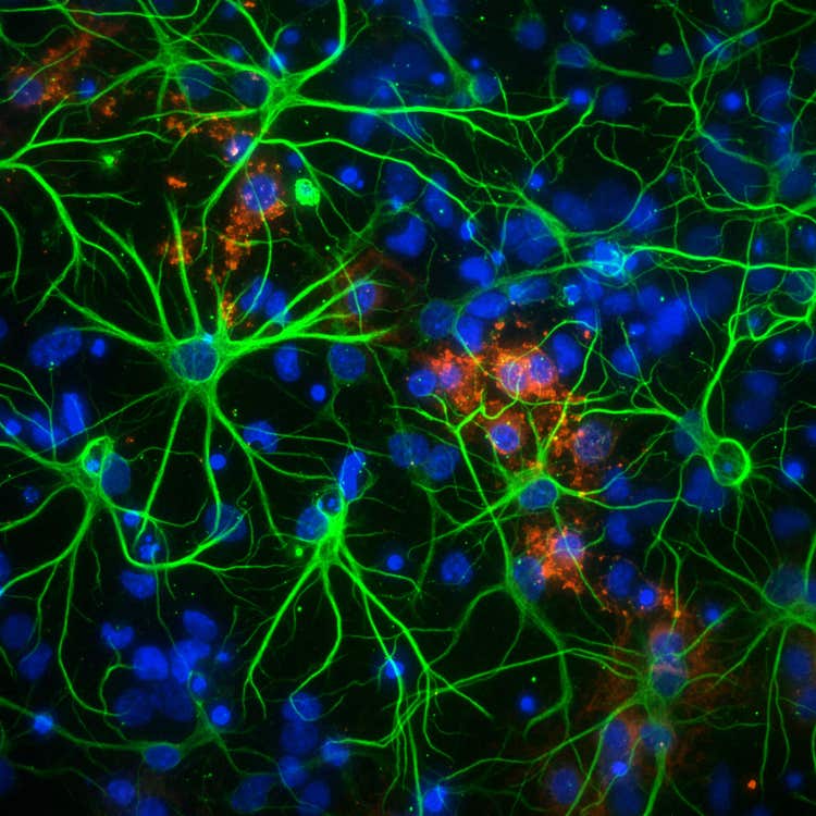
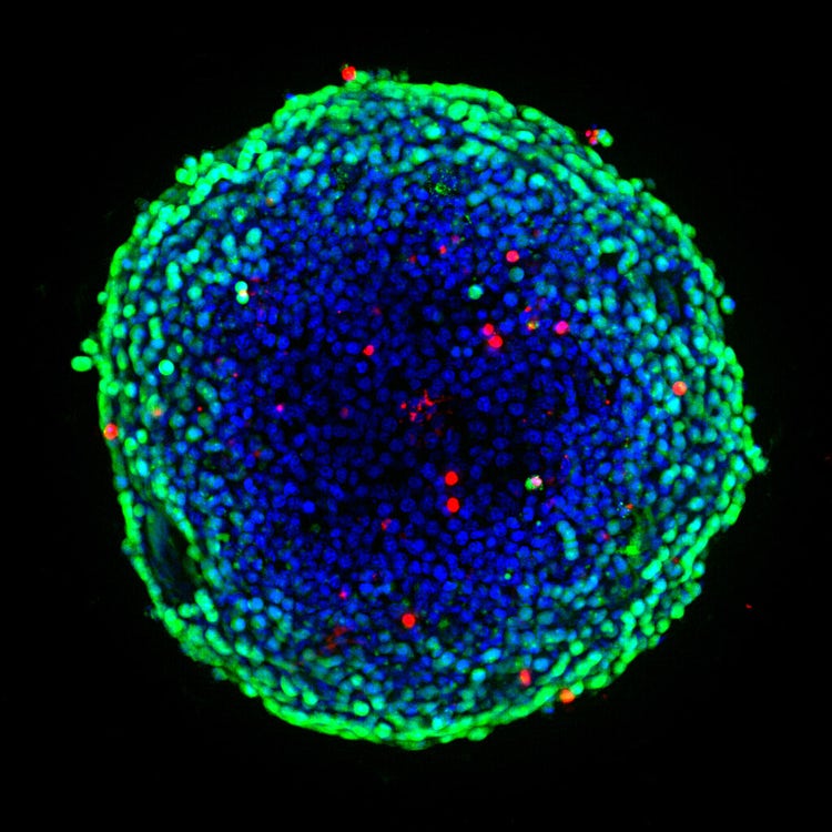
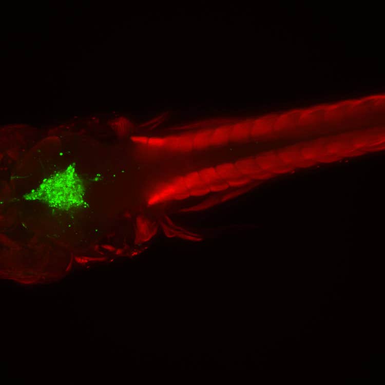
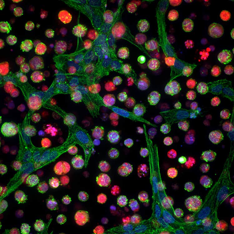
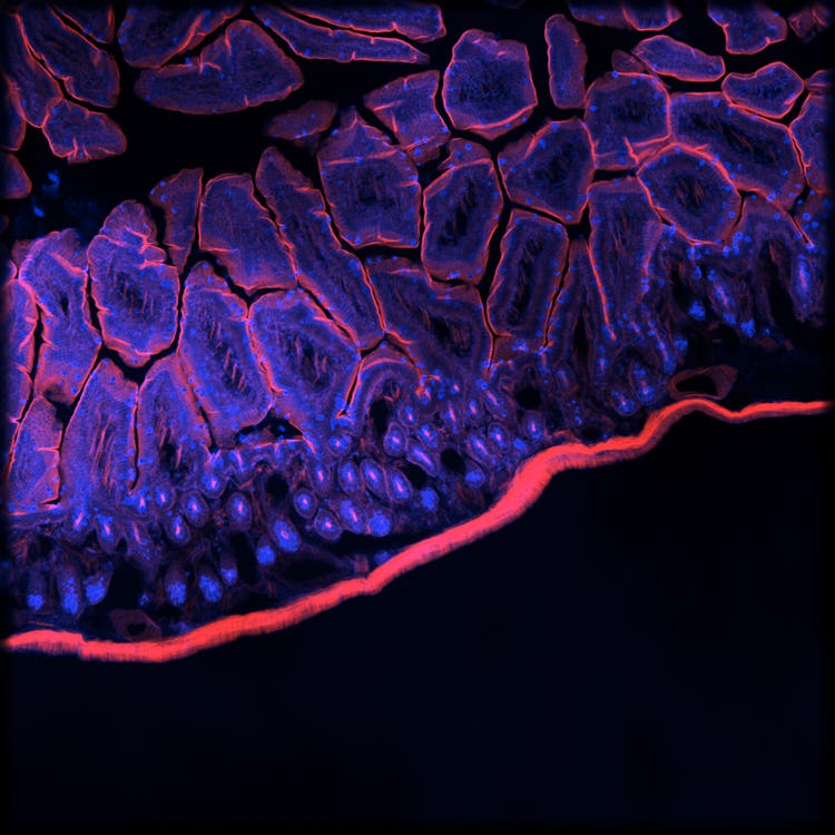
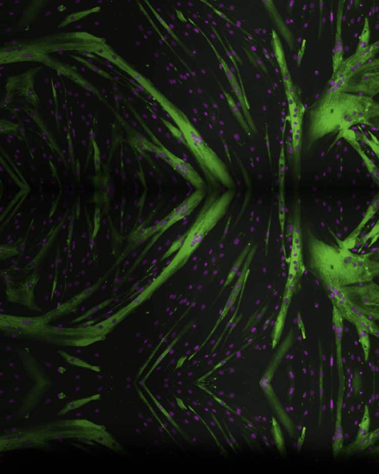
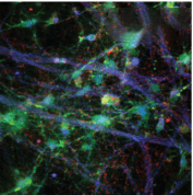
세포 이미지 갤러리
Micro Confocal High-Content Imaging 시스템
ImageXpress High-content imaging 솔루션
ImageXpress Micro Confocal Imaging 시스템
ImageXpress Micro Imaging 시스템
Micro Confocal Imaging 시스템
†고객이 제공한 샘플을 사용하여 개발 중 데이터 및 이미지를 획득했습니다. 결과에는 차이가 있을 수 있습니다. 주요 기능의 가격, 배송 기간, 사양은 상호간에 동의한 기술적 요구 사항에 따라 달라집니다. 솔루션 요구 사항으로 인해 표준 성능이 조정될 수 있습니다.
최신 자료
제품 응용 분야
고객 사례 소개

성공 사례
Visikol 연구팀은 ImageXpress Micro Confocal High-Content Imaging 시스템과 MetaXpress 소프트웨어를 사용하여 NASH 및 NAFLD 질병 진행 과정을 시뮬레이션하는 3D 세포 배양 모델을 개발합니다.
유연한 고성능 Imaging 솔루션을 사용한 연구 확장
Molecular Devices는 연구 요구 사항을 충족하고 환경 제어 하에서 세포 건강 상태 운동 기능을 모니터링하기 위해 행잉 드롭(hanging drop) 및 원형 또는 평면 바닥 플레이트 등 다양한 샘플 형식으로부터 이미지를 손쉽게 포착할 수 있도록 ImageXpress Micro Confocal High-Content Imaging 시스템을 위한 유연한 옵션을 제공합니다. 30년 이상의 이미징 전문 지식을 보유한 당사에서 최상의 assay용 이미지를 보장하기 위한 적합한 옵션을 선택하시도록 도와 드릴 수 있습니다.
표준 하드웨어 옵션

Water Immersion Objective
20X, 40X 및 60X Water Immersion Objective는 획득 중에 기하학적 정확성을 개선하고 빛 굴절을 줄여 짧은 노출 시간에 더 밝은 세기를 냅니다.

Transmitted Light 타워
당사의 Transmitted Light 타워는 쉽게 보거나 Background에서 분리될 수 있는 미염색(Unstained) 세포에 대한 High Contrast 이미지의 획득을 지원합니다.

EC(Environmental Control)
Environmental Control은 장기간 Live cell/ Time-lapse 분석 시, 온도를 제어하고 수분 증발을 최소화합니다.

온 보드 로봇 Fluidics 장치
통합된 Fluidics 장치는 화합물 첨가, 웰 세척 및 배지 교환이 필요한 assay 워크플로를 자동화합니다.
맞춤형 옵션
Molecular Devices는 아래에 설명된 기능을 포함한 맞춤형 소프트웨어 및 하드웨어가 ImageXpress Micro Confocal High-Content Imaging 시스템에 포함되도록 성공적으로 조절할 수 있으며, 배양기, 액체 처리기, 로봇 등과 같은 기타 실험실 구성요소를 통합하여 완전히 자동화된 작업셀을 구현할 수 있습니다. 생명과학 분야에서 30년 이상의 경험과 더불어, 품질이 우수한 제품을 납품하고 전 세계 지원 서비스를 제공하고 있으므로 신뢰하실 수 있습니다.
판매는 www.moleculardevices.com/custom-products-purchase-terms에서 확인 가능한 당사의 맞춤형 제품 구매 약관에 따름

고강도 레이저
5채널 또는 7채널 고강도 레이저를 통해 실험 역량을 확장합니다.

실시간 용량 반응
자동 피펫터를 사용하면 화합물을 추가하는 동시에 초당 >100프레임에서 실시간 스트리밍을 할 수 있습니다.

심부 조직 투과, 공초점 디스크 모듈
심부 조직 투과, 공초점 디스크 모듈은 혼선을 줄여 아웃포커스 광억제를 개선하고 조직에 더 깊이 투과합니다.

턴키, High-Throughput 장기 동역학
일정한 온도, O2(저산소증), CO2 및 습도 조건을 유지하면서도 장기간에 걸쳐 다수의 플레이트를 일정 수립 및 이미징할 수 있습니다. Live Cell 워크어웨이 용량을 플레이트 200개 이상으로 확장할 수 있습니다.

로봇 자동화 확장
처리량을 증가시키고, 사람의 실수를 없애고, 멸균성을 유지하고, 일관된 샘플 처리를 달성할 수 있습니다. 모듈식 자동화 설계—구성품을 모듈 단위로 추가할 수 있으며 업그레이드가 가능합니다.
고강도 레이저
고출력 레이저 Excitation으로 노출 시간을 최대 75%까지 줄일 수 있습니다.† 레이저 광원은 출력이 400~1,000mW/채널인 5채널 또는 7채널 광원으로 사용할 수 있습니다. 7채널 레이저 광원은 근적외선을 포함하며 다중 요구 사항이 있는 고객에게 적합합니다.
- 신호-소음 비율이 더 높은 보다 선명한 이미지 획득
- 스캔 속도가 최대 2배†까지 향상되어 노출 시간이 크게 감소합니다.
- CFP 및 YFP용 레이저를 사용하여 FRET 실험 실행
심부 조직 투과, 공초점 디스크 모듈
레이저 광원과 결합된 심부 조직 투과에 특화된 공초점 디스크 모듈은 심부 조직 투과를 위한 빛 투과를 개선하고, 두꺼운 조직 표본의 이미지를 더 선명하게 만들어 Imaging 해상도를 개선할 수 있습니다†.
- 아웃포커스 광선의 억제 개선
- 흐릿함 감소(핀홀 혼선)
- 두꺼운 조직 샘플에 대한 더 깊은 투과로 더욱 선명한 이미지


