
Water Immersion Objective
Water Immersion 기술이란?
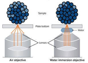
Water Immersion은 High Content Imaging에서 대물렌즈(배율)와 표본 사이에 물의 층을 자동으로 일관되게 배치하는 기술입니다.
많은 세포 Imaging 시스템에서 대물렌즈와 표본 사이에 공기를 배치하도록 설계되어 있습니다. 3D Imaging에 공기 대물렌즈를 사용하는 경우, 시료에서 방출되는 형광의 상당한 부분이 대물렌즈에서 굴절되어 소실됩니다. 이는 시료에서 수집된 신호를 감소시키며 시료가 왜곡되어 나타나도록 합니다.
오일 대물렌즈는 시료에서 더 많은 빛을 수집하여 신호 강도를 높이고 이미지 해상도를 향상시켰기 때문에 3D 공초점 Imaging의 최적 표준이었습니다. 그러나 오일을 자동화된 방식으로 다루기 힘들며 이 대물렌즈에서도 대규모 3D 시료를 Imaging할 때 약간의 왜곡이 생기므로 오일 대물렌즈에는 여전히 문제가 있습니다.
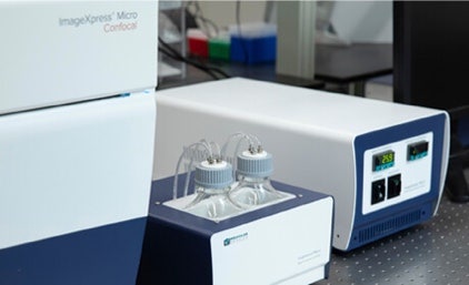
ImageXpress Micro Confocal 시스템용 Water Immersion Objective
과학자들은 복잡한 모델을 Screening하고 더 우수한 데이터 품질을 획득하는 데에 어려움을 자주 겪습니다. ImageXpress® Micro Confocal High-Content Imaging 시스템에서 Water Immersion Objective를 사용하게 되면, 신호를 최대 4배로 증가시킬 수 있으며, Z 해상도를 개선하고, 광학적 장치 수차를 감소시켜 더 선명하고 생생한 이미지를 볼 수 있습니다.
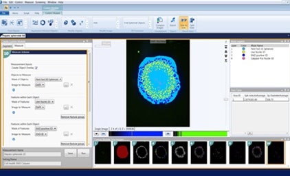
3D 이미지 분석 실험과정을 다루기 위해, 우리의 MetaXpress® 3D 분석 소프트웨어 모듈은 처리량이나 데이터 품질의 저하 없이 단일 인터페이스 안에 더 많은 표현형 데이터를 생성할 수 있도록 하여 과학자들이 스스로의 발견에 더 확신을 가질 수 있게 하였습니다.
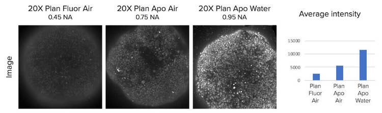
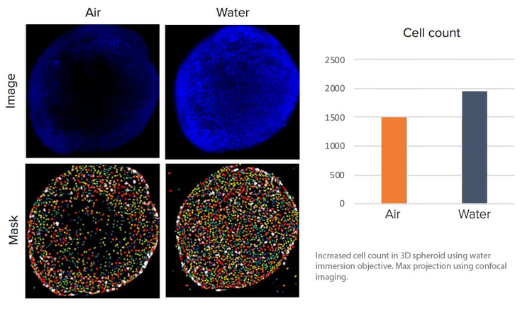
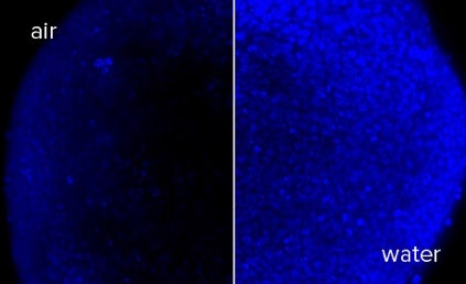
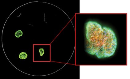
Water Immersion Objective를 사용한 3D Assay의 평균 강도 증가. 50 ms 노출에서 스페로이드의 핵, 공초점 Imaging을 이용한 최대 투사.
†고객이 제공한 샘플을 사용하여 개발 중 데이터 및 이미지를 획득했습니다. 결과에는 차이가 있을 수 있습니다.
보다 깊이 있게 더 많은 표현형 데이터를 포착하기 위한 감도를 확보합니다.
Water Immersion Objective는 신호를 최대 4X†까지 증가시킬 수 있으므로, 과학자들이 적은 노출 시간에도 3D 및 두꺼운 조직 샘플을 더 자세히 볼 수 있게 합니다.

최대 4배 더 많은 깊이 있는 신호 확보
z-resolution를 개선하고 광학적 수차를 줄여 3D 샘플을 좀 더 정확하게 재구성할 수 있습니다.
더욱 선명하고 깨끗한 이미지 확보
더 나은 광 수집으로 더 짧은 노출 시간에 보다 밝은 세기의 이미지를 얻을 수 있으므로, 데이터의 품질 개선에 기여합니다.
자신감 있게 샘플 실행
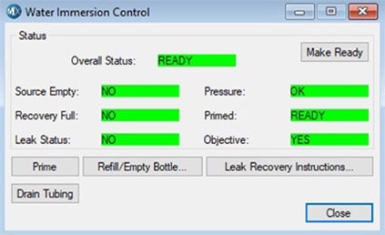
MetaXpress 소프트웨어에는 물 컨트롤러 시스템의 시스템 상태를 표시하기 위해 통합된 사용자 직관적인 센서와 경보가 포함되어 샘플 실행 시 안심할 수 있습니다.
고처리량에서 3D 샘플 획득
Water Immersion Objective가 포함된 ImageXpress Micro Confocal 시스템은 획득 중에 물을 자동으로 보충하기 때문에 전체 Microplate 샘플을 High-throughput으로 획득할 수 있습니다.
3D 구조의 보기 및 정량화 간소화
스페로이드, 미세 조직, 세포를 3D 매트릭스로, 그리고 z 평면의 스택으로 획득한 작은 유기체를 분석합니다. MetaXpress 3D 분석 모듈을 활용하여 부피, XYZ 위치, 인접 대물과의 거리, 직경, 깊이, 다양한 세기 측정 수치, 질감 또는 대물의 수를 평가할 수 있습니다.
이미지 획득 시간 단축
관심 대상이나 드문 Event를 찾기 위한 낮은 배율의 QuickID 표적 획득 이미지는 필요한 파장, Z-stack 또는 시점으로 고배율 이미지를 자동 획득합니다. 이 분석 프로세스는 선택한 표적을 고배율로 획득하여 획득 시간을 단축하고 이미징 저장 요구사항을 줄입니다.
†고객이 제공한 샘플을 사용하여 개발 중 데이터 및 이미지를 획득했습니다. 결과에는 차이가 있을 수 있습니다.