생체모사사칩 플랫폼에서 3D High-Content Imaging 및 분석 가능
질병 모델링 및 약물 Screening을 위한 인체의 생물학적 환경을 모방하는 능력이 있고, 이를 미세한 크기의 시스템에서 수행한다고 상상해 보세요. With the development of organ-on-a-chip models, this is no longer implausible. Integrating microfluidics with cells and growth factors, scientists can now generate biologically relevant models emulating the physiological and mechanical behaviors of the actual tissue.
With successful implementation, organ-on-a-chip models can save significant time in drug discovery, enabling faster and more accurate assessment for pharmacokinetics, efficacy and cytotoxicity, and personalized medicine research.
In a not-so-distant future, we will be less dependent on animal models for drug development and toxicity assessment tests as they more accurately mimic in vivo conditions.
These microscale organ systems are designed to represent the intricacies of human biology better. However, their complexity presents challenges around imaging at scale to obtain quantitative and reproducible results.
The groundbreaking collaboration between Molecular Devices and MIMETAS, a global leader in organ-on-a-chip models, can help us demonstrate how to overcome these 3D high-content imaging challenges.
What is an organ-on-a-chip?
Organs-on-chips are microfluidics-based cell culture devices that are the size of a memory stick and can recapitulate the complex structures and processes of human organs. They consist of a flexible and translucent biopolymer with tiny hollow channels containing living human cells.
By specifically arranging and compartmentalizing different cell types, one can simulate and control the interactions between other tissue cells in the organ. A porous membrane usually separates the microchannels to enable intercellular crosstalk, such as the interactions between endothelial and epithelial cells.
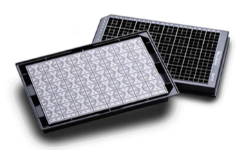
OrganoPlate: MIMETAS’ cutting-edge solution to tissue modeling
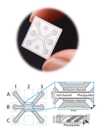
Founded in 2014 in Leiden, the Netherlands, MIMETAS is a pioneer of organ-on-a-chip models enabling the study of human disease models. Their key innovation, OrganoPlate®, is a microfluidics-based platform that supports up to 96 tissue models on a single plate.
OrganoPlate owes its versatility and simplicity to PhaseGuide™ technology. Unlike conventional organs-on-chips, the patented liquid handling technology enables the membrane-free formation of extracellular matrices in 3D. This allows the free movement and interactions of cells and the introduction of proteins and co-culture compounds.
The microfluidics networks in OrganoPlate facilitate a continuous medium flow without external pumps or tubing. The movement of nutrients, oxygen, growth factors, and co-culture conditions is mediated by a gravity-driven technology. This results in continuous perfusion that mimics blood flow. These factors make OrganoPlate an excellent source for studying permeability, migration, outgrowth, invasion, angiogenesis, and complex cell-cell interactions.
3D high-content imaging solutions for organ-on-a-chip
Since organ-on-a-chip models are compatible with various assays, from immunostaining to viability to qPCR, the monitoring and analysis of results are often more complex than 2D in vitro models.
The good news is, OrganoPlate contains a microscopy-grade glass bottom that makes it the only organ-on-a-chip model suitable for high-throughput and high-content imaging.
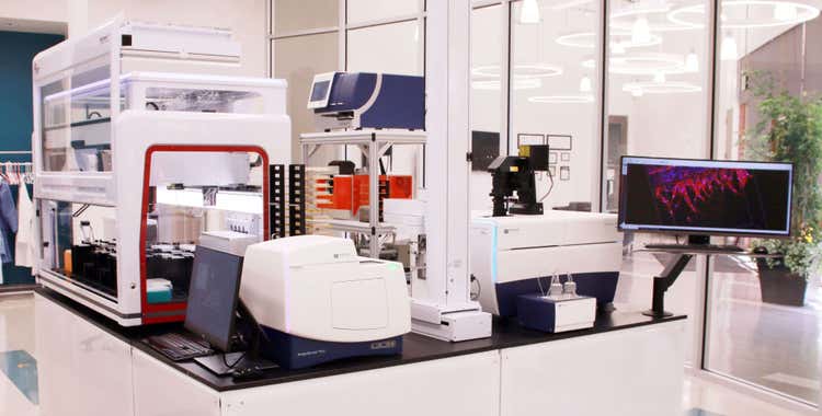
MIMETAS has collaborated with Molecular Devices to enhance their 3D high-content imaging skills and equipment. One of the recent products of this collaboration is the installation of Molecular Devices’ new ImageXpress® Micro Confocal High-Content Imaging System. Taking it one step further, we have also been offering personalized training to MIMETAS’ customers. Researchers can learn how to design and conduct organ-on-a-chip experiments on OrganoPlate and image the 3D cell cultures using ImageXpress Micro Confocal.
Let’s now look at how this applies to specific tissue models.
혈관생성
Angiogenesis involves the formation of new blood vessels, as well as the regeneration of damaged ones. It is not only an integral part of tissue growth, development, and repair, but a common phenomenon in tumor invasion and metastasis. Therefore, successful imaging of angiogenic mechanisms can open doors to anti-angiogenic cancer drug discovery. However, current in vitro assays provide limited insight and fail to illustrate endothelial sprouting and vessel growth.
The OrganoPlate platform provides researchers with a more informed model to quantify and visualize angiogenic sprout number and size, and angiogenic markers for proliferation and expression.
During the first angiogenesis study conducted by MIMETAS with Molecular Devices, OrganoPlate 3-lane was used to develop the angiogenesis model. Each of the 40 chips in OrganoPlate consisted of three channels, with the following content from top to bottom: endothelial cells, collagen-I extracellular matrix (ECM) gel, and angiogenic factors. Then, as depicted in the application note, 3D Image Analysis and Characterization of Angiogenesis in Organ-on-a-Chip Model, the formation of angiogenic sprouts was monitored over the next four days.
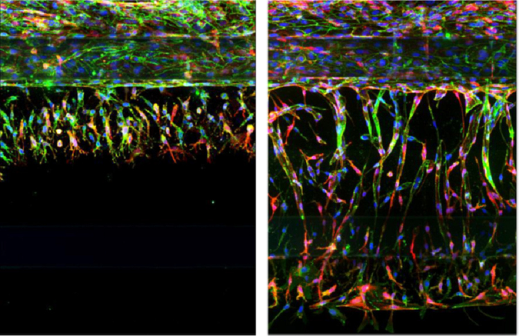
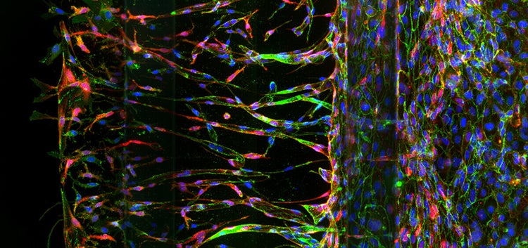
Day 1 (left) and day 4 (right) of angiogenic sprouts in OrganoPlate with 20X magnification.
The nuclei, the cell adhesion molecule VE-cadherin, and actin filaments of endothelial cells were differentially stained. Using the confocal mode of ImageXpress Micro Confocal High-Content Imaging System, researchers could visualize sprouts after 1 day and 4 days in culture. Further analysis of these images was carried out with the Custom Module Editor (CME) in MetaXpress® High-Content Image Acquisition and Analysis Software. This analysis provided valuable insights into the time-dependent growth of the vessels by revealing various factors, such as the total number of sprouts, number of nuclei per sprouts, volume, intensity, and distances between sprouts.
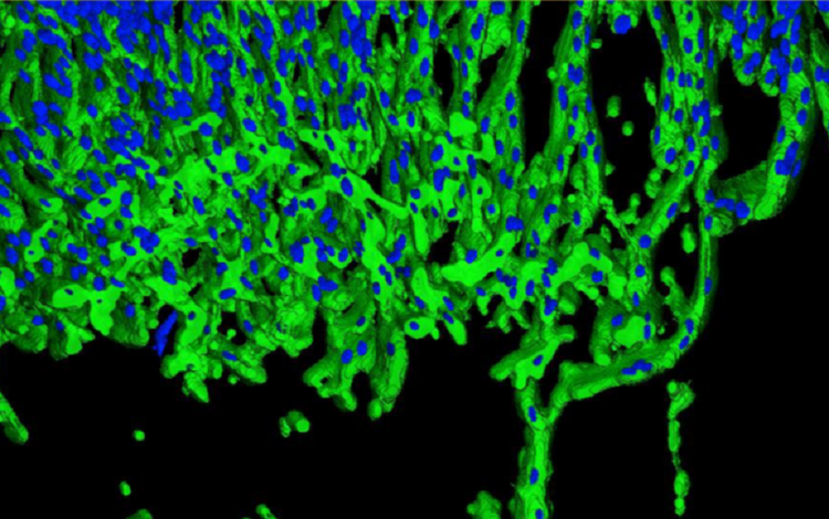
3D analysis of angiogenic sprouts (green) and nuclei (blue) using MetaXpress® High-Content Image Acquisition and Analysis Software.
Neurite Outgrowth
Drug development for neurodegenerative diseases requires a rigorous understanding of the connections between neurons. These connections, mediated by the extended neural structures on cells called axons and dendrites, are defined as neurites. Therefore, neurite outgrowth is a significant indicator of neural connectivity or disruptions within it.
Neurite outgrowth assays can help visualize and quantitatively analyze intracellular and extracellular signals engaged in neuronal degeneration, but this requires physiologically relevant 3D tissue models.
To investigate the inhibition of neurite outgrowth, researchers from MIMETAS used the OrganoPlate platform to develop microfluidics-based and sustainable neuronal cultures. As shown in High-Content Assay for Morphological Characterization of 3D Neuronal Networks in a Microfluidic Platform, human iPSC-derived neurons were treated with compounds, which inhibited neurite outgrowth and were differentially stained with three different dyes.
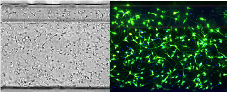
iCell neurons in OrganoPlate capillary well for 72 hours (left) and live cells stained with three dyes examined with ImageXpress® Micro Confocal High-Content Imaging System (right).
The ImageXpress Micro Confocal High-Content Imaging System aided with the assessment of neural viability, as well as morphological changes in neurons. 3D analysis with MetaXpress software returned quantitative data, such as the number of neurites, neuron volume, number of nuclei, and number of neurite branching.
The results proved that the combination of MIMETAS’ OrganoPlate and Molecular Devices’ high-content imaging systems can be used for assessing neurological drug efficacy and toxicity.
Get started with 3D human tissue models and imaging
Efficient implementation of organ-on-a-chip systems requires a thorough understanding of the working mechanism. Luckily, all the information you need is one click away.
In, Getting Started with 3D Human Tissue and Imaging, Chiwan Chiang (Field Application Scientist, MIMETAS) and Oksana Sirenko, PhD (Sr. Applications Scientist, Molecular Devices) demonstrate how to get powerful new insights from these models quickly, easily, and affordably.
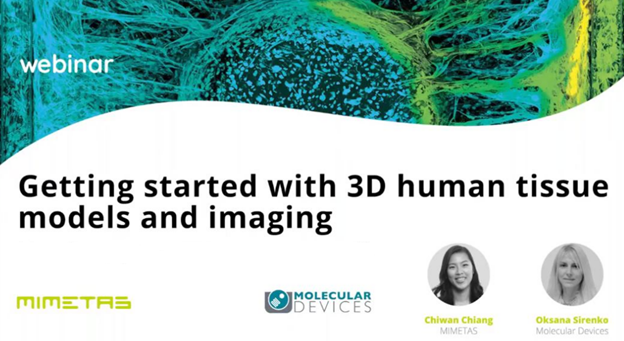
Dive into the setup of 3D organotypic tissue models in the OrganoPlate, and discover the role high-content imaging plays in organ-on-a-chip studies. Presenters discuss the benefits of human tissue modeling with two case studies. Case study one, gut-on-a-chip, involves the caco-2 gut model to assess barrier integrity. The second case study, angiogenesis, pertains to angiogenic sprouting in HUVEC micro vessels.
Need help with your organ-on-a-chip workflow?
Find out more about our Organoid Innovation Center and extensive portfolio of solutions.