Molecular Devices, LLC
  •   美谷分子仪器(上海)有限公司
    咨询服务热线:400-821-3787
  •   0 Cart
  • Store

  •  채용
  •  언어  
    • English
    • Deutsch
    • 中文
    • 日本語
    • Français
    • 한국어
    • Italiano
    • Español
Molecular Devices, LLC
  • 제품
    • 제품

      새로운 DispenCell™ Single-Cell Dispenser 기술은 단일 세포주를 3배 더 빠르고 저렴한 비용으로 분리합니다.

      • 쉽고 직관적인 설정
      • 클론형성능과 추적 가능성의 즉각적인 증거
      • 고유한 기술로 세포 샘플을 부드럽게 처리
      • 벤치탑 크기  
        설계
      • 특허받은  
        일회용 팁
      DispenCell™ Single-Cell Dispenser 기술

      최적화된 3D 조직과 오가노이드 실험과정을 위한 BioAssemblyBot의 6축 로봇 팔을 갖춘 자동화된 High-Content Screening 솔루션

      BioAssemblyBot의 6축 로봇 팔

    • Microplate Reader
      SpectraMax Mini Multi-Mode Microplate Reader
      Multi-Mode Reader
      • SpectraMax i3x
      • SpectraMax iD3/iD5
      • SpectraMax M 시리즈
      • FlexStation 3
      • SpectraMax Mini
      SpectraMax ABS 마이크로플레이트
      흡광(Absorbance) 리더기
      • SpectraMax ABS/ABS Plus
      • SpectraMax VersaMax
      • SpectraMax QuickDrop
      • CMax Plus
      Fluorescence Reader
      Fluorescence Reader
      • SpectraMax Gemini
      SpectraMax Luminescence
      발광(Luminescence) 리더기
      • SpectraMax L


      MultiWash+ Washer
      Stacker 및 Washer
      • StakMax Stacker
      • AquaMax Washer
      • MultiWash+ Washer
      • MultiWash–C 微孔板洗板机
      SoftMax Pro 데이터 획득
      분석 소프트웨어
      • SoftMax Pro 소프트웨어
      • SoftMax Pro GxP Software
      GxP 솔루션
      GxP Compliance Solution
      • SoftMax Pro GxP Software
      • 소프트웨어 설치와 Validation 서비스
      • IQ/OQ/PM 서비스
      • SpectraTest Validation Plate
      실험실 자동화와 맞춤화
      실험실 자동화와 맞춤화
      • 플레이트 기반 High-Throughput Assay를 위한 실험실 자동화
    • 세포 Imaging 시스템
      ImageXpress Pico Automated Cell Imaging System
      Automated Cell Imaging Systems
      • ImageXpress Pico
      • ImageXpress Nano
      High-Content Imaging
      High-Content Imaging
      • ImageXpress Confocal HT.ai
      • ImageXpress Micro Confocal
      • ImageXpress Micro 4
      StratoMineR 분석
      Acquisition & Analysis Software
      • IN Carta
      • StratoMineR
      • MetaXpress
      • CellReporterXpress
      • MetaMorph
      실험실 자동화와 맞춤화
      실험실 자동화와 맞춤화
      • High-Throughput, High-Content Screening(HCS)을 위한 실험실 자동화
      • BioAssemblyBot 400 Bioprinter 자동화 HCS 솔루션
    • 클론 스크리닝
      Clone Pix 시리즈
      포유류 콜로니 피킹
      • ClonePix 2
      QPix 미생물 콜로니 피커
      미생물 콜로니 피킹
      • QPix 420
      • QPix 450/460
      • QPix HT
      CloneSelect Imager FL
      단일 세포 Imaging
      • CloneSelect Imager
      • CloneSelect Imager FL
      DispenCell Single-Cell Dispenser
      단일 세포 분리
      • DispenCell Single-Cell Dispenser
      실험실 자동화
      실험실 자동화와 맞춤화
      • High-Throughput 클론 스크리닝을 위한 실험실 자동화
      CloneMedia와 XP Media
      배지 및 시약
      Clone Screening Assay kit
      Clone Screening Assay kit
    • FLIPR Penta
      FLIPR Penta
      FLIPR Penta
      • FLIPR Penta High-Throughput 세포 스크리닝 시스템
      Screenworks
      분석 소프트웨어
      • ScreenWorks 소프트웨어
      • Peak Pro 2 소프트웨어 모듈
      Flipr Assay 키트
      FLIPR Assay 키트
      • 칼슘 Assay 키트
      • 칼륨 분석 키트
      • 막전위 분석키트
      • EarlyTox 심장 독성 키트
    • Axon Patch-Clamp
      투명
      Amplifier
      • Axopatch 200B 콘덴서
      • MultiClamp 700B
      • Axoclamp 900A
      투명
      Digitizer
      • Axon Digidata 1550B Low
      투명
      분석 소프트웨어
      • pCLAMP 11 소프트웨어 제품군
    • 기타
      Threshold Immunoassay System
      Threshold Immunoassay System
      Geneppix 미세배열 스캐너
      Genepix 미세배열 스캐너
      Imagexpress Micro Xls
      Imagexpress Micro xls
      인증 리퍼브
      인증 리퍼브
      IDBS 솔루션
      IDBS R&D 클라우드 솔루션
    • 분석 시약(assay kit)
      심장 독성
      • EarlyTox 심장 독성 키트
      Cell viability
      • EarlyTox Cell Integrity Kit
      • EarlyTox Cell Viability Assay Kit
      DNA 정량 분석
      • Spectramax Quant dsDNA Assay 키트
      Elisa, western blot
      • CatchPoint SimpleStep ELISA 키트
      • ScanLater Western Blot Assay 키트
      gpcr
      • FLIPR 칼슘 Assay 키트
      • Fura-2 QBT 칼슘 키트
      • CatchPoint cAMP 형광 Assay 키트
      • CatchPoint cGMP 형광 Assay 키트
      이온 통로
      • FLIPR 칼륨 Assay 키트
      • FLIPR 막전위 Assay 키트
      IGG 정량분석
      • ValitaTiter
      • CloneDetect
      Reporter gene
      • Spectramax Glo Steady-Luc Reporter Assay Kit
      • Spectramax DuoLuc Reporter Assay Kit
      Transporter
      • QBT Fatty Acid Uptake Assay Kit
      • 신경전달물질 수송체 흡수 Assay 키트
      기타
      • Contamination Detection
      • Enzyme - IMAP Assay
    • 액세서리 및 소모품
      Microplate Reader
      • 384 Well SBS
      • 384 Well High Sample Recovery Plate
      • Deep-well Plate
      • Low Profile Microplate
      • SpectraDrop Micro-Volume Microplate
      • SpectraMax Injection cartridge with SmartInject Technology
      • SpectraMax MiniMax 300
        Imaging Cytometer
      • Western Blot Cartridge
      • 96孔微孔板
      클론 스크리닝
      • Adjustable Petri Dish and Microplate Holder
      • Bioassay QTrays
      • Calibead
      • 캡 매트 및 뚜껑
      • 크로마 필터
      • 세정 및 살균 솔루션
      • CloneSelect Single-Cell Printer Cartridge
      • QPix 핀 및 헤드
      • QReps 레플리케이터
      Axon Patch-Clamp
      • Soft Panel Amplifier Control
      Spectra Img
  • 연구 분야
    • 응용 분야

      Molecular Devices, Cellesce 인수로 환자 유래 오가노이드 특허 기술 추가

      2022년 12월 6일

      • Cellesce의 동종 업계 최초 기술로 대규모 약물 Screening을 위한 일관적인 환자 유래 오가노이드를 생성합니다.
      • 인수를 통해 Molecular Devices의 3D 생물학 솔루션 혁신 기업으로서의 입지를 강화합니다.
      • 결합된 전문 지식으로 신약 개발을 위해 생리학적으로 연관된 세포 모델의 채택을 업계에서 가속화할 것입니다.


      OIC 방문하기

      언론 보도 읽기

      Cellsce
      응용 분야를 위한 Spectra
    • 코로나바이러스 (COVID-19)
      코로나19
      코로나19 연구
      관련 솔루션
      코로나19
      코로나19 관련
      새로운 뉴스
      코로나19
      백신 개발 워크플로
      감염병 연구 응용 분야
      백신 연구
      연구 응용 분야
    • 연구분야 (Stem Cell, Cancer)
      투명
      3D Cell Model
      투명
      암 연구 솔루션
      투명
      Cell Line Development
      투명
      신약 개발
      투명
      식품 및 음료
      투명
      유전자 편집(CRISPR/Cas9)
      투명
      오가노이드 연구
      투명
      줄기세포 연구
      투명
      독성학
    • Microplate Reader
      투명
      세포 건강 상태 (Cell Health)
      투명
      Cellular Signaling
      투명
      ELISA
      투명
      미생물학과 오염물질
      투명
      핵산(DNA/RNA) 측정과 분석
      투명
      단백질 측정, 정량분석, 분석
      투명
      관련 분석법: 측정 모드
      • 흡광(Absorbance)
      • 형광(Fluorescence)
      • 형광 편광
      • 발광(Luminescence)
      • TRF, TR-FRET 및 HTRF
      • Western Blot
    • 세포 Imaging 시스템
      투명
      Cell Counting
      투명
      세포 이미징 및 분석
      투명
      세포 Migration Assay
      투명
      세포 염색
      투명
      Live Cell Imaging
      투명
      Neurite Outgrowth
      투명
      장기 칩
      투명
      오가노이드
      투명
      Spheroids
    • 클론 스크리닝
      투명
      Cell line development 실험과정
      투명
      단클론항체(mAb)
      • 하이브리도마
      • 파지 디스플레이
      • 단일클론항체 생산
      투명
      Monoclonality
      투명
      합성 생물학
    • FLIPR Penta
      투명
      GPCR(G protein-coupled receptor)
      투명
      이온 채널
      투명
      Cardiotoxicity(심장독성)
    • Axon Patch-Clamp
      투명
      전기생리학(Patch Clamp)
  • 자료
    • 자료
    • 관련 자료 검색
      메뉴 자료 아이콘/응용 분야 노트
      Application Note
      메뉴 자료 아이콘/레퍼런스
      레퍼런스
      Ebook 아이콘
      eBook
      메뉴 자료 아이콘/Scientific Poster
      Scientific Poster
      메뉴 자료 아이콘/튜토리얼 및 영상
      동영상 및 웨비나

      검색

    • 블로그 – 실험실 노트
      스페이서
      Assay에 대한 고객 사례…
      스페이서
      3D organoids and…
      How 3D Cell Models Will Shape the Future of Drug Discovery
      2023년 3월 7일 Target discovery and drug development rely heavily on 2D cell and animal models to decipher efficacy and toxic effect of drug candidates. Yet, 90% of candidates fail to…
      더 알아보기  
    • 고객 사례 소개

      다른 연구자들이 제품과 솔루션을
      어떻게 사용했는지 확인해보세요.

    • 혁신 기술
      투명
      AI, 머신러닝 및 딥러닝
      투명
      AgileOptix 스피닝 디스크 기술
      투명
      자동 초점
      투명
      디지털 공초점 옵션
      투명
      고함량 Screening
      투명
      HumSilencer
      투명
      레이저 조명
      투명
      QuickID 표적 이미지 획득
    • 관련 분석법
      투명
      흡광(Absorbance)
      투명
      전기생리학
      투명
      형광(Fluorescence)
      투명
      형광 편광(FP)
      투명
      발광(Luminescence)
      투명
      TRF, TR-FRET 및 HTRF
      투명
      Water Immersion Objective
      투명
      Western Blot
    • 동영상 갤러리
      투명
      Microplate Reader
      투명
      세포 Imaging 시스템
      투명
      Flipr 시스템
      투명
      클론 스크리닝
      투명
      Axon Patch-Clamp
      투명
      주문형 웨비나
  • 서비스 및 지원
    • 서비스 및 지원
    • 개요
      Spectra 로고
      SpectraNet 고객 관리 포털
      GxP 컴플라이언스
      GxP 컴플라이언스 솔루션
      실험실 자동화와 맞춤화
      실험실 자동화와 맞춤화
      전문 서비스
      전문 서비스

      기술 지원

      미국 본사  
      +1 800-635-5577  
      월~금, 오전 7시~오후 5시 PST

      유럽  
      +44-118-944-8000  
      월~금, 오전 8시~오후 5시 GMT

      내가 있는 지역의 담당자 확인하기

      SpectraNet 고객 포털  

    • 고객 포털 - Spectranet
      Spectranet

      INTRODUCING OUR NEW CUSTOMERCARE PORTAL

      SpectraNet is an intuitive, simple-to-use, self-service customer portal providing a new level of experience available 24/7.

      Create your account today to get full access to integrated content and world-class customer service.

      REGISTER LOGIN
      지원 티켓 신청
      지원 티켓 신청하기
      자료 창고
      지식 기반
      제품 등록
      제품 등록하기
      기술 자료
      기술 자료
    • GxP 컴플라이언스 솔루션
      GxP Softmax Pro GxP 소프트트웨어
      SOFTMAX PRO GXP 소프트웨어
      GxP 소프트웨어 설치
      소프트웨어 설치와 Validation 서비스
      GxP Spectratestt Validation Plate 재인증
      SPECTRATEST Validation Plate
      IQ OQ 서비스
      IQ/OQ/PM 서비스
    • 실험실 자동화
      실험실 자동화와 맞춤화
      실험실 자동화와 맞춤화
      High Content Screening HCS
      High-Throughput, High-Content Screening
      • BioAssemblyBot 400 Bioprinter 자동화 HCS 솔루션
      플레이트 기반 High-Throughput Assay
      플레이트 기반 High-Throughput Assay
      High-Throughput 클론 스크리닝
      High-Throughput 클론 스크리닝
    • 전문 서비스
      전문 서비스
      전문 서비스
      서비스 플랜
      서비스 플랜
  • 회사
    • 회사

      Molecular Devices, 오스트리아의 글로벌 R&D 허브 확장

      2022년 10월 12일        
      더 큰 부지는 Cell Line Development, 오가노이드 개발, 신약 개발을 개선하기 위한 Screening 솔루션을 발전시키기 위한 협업 공간인 잘츠부르크의 오가노이드 혁신 센터의 미래 기지가 될 것입니다.


      OIC 방문하기

      언론 보도 읽기

      Austrian Research & Development Center의 리본 커팅 기념식
      응용 분야를 위한 Spectra
    • 회사 소개

      Molecular Devices는 실리콘 밸리를 기반으로 제약 및 연구의 스크리닝과 효과적인 분석이 가능한 솔루션을 40년에 걸쳐 개발하고 공급해 왔습니다.

    • 리더십

      당사의 다양한 경험, 비즈니스 인사이트 및 공동의 목표의식은 직원들이 잠재력을 최대한 발휘하도록 독려하기 위한 당사의 일상적인 결정에 추진력을 부여합니다.

      리더십

    • 채용

      당사의 팀 중심 기업 문화는 생각과 관점의 다양성과 강력한 신뢰 관계를 보장합니다.

    • 뉴스룸
      투명
      뉴스
      투명
      보도 내용
      Silver Sponsor Molecular Devices at Society for Laboratory Automation and Screening 2023 International Conference and Exhibition
      Feb 22, 2023 Showcasing new industry collaborations, automated technology, and workflow innovations that span 3D biology, cell line development, and drug…
      Read more  
    • 이벤트
      Focus on Microscopy (FOM)
      Conference | Europe | Porto, Portugal, Europe– Apr 02 – Apr 5, 2023 FOM2023 continues a long-standing (since 1988), yearly conference series on the latest innovations and developments in (optical) microscopy and their…
      Read more  
      Imaging User Meeting 2023
      Conference | Europe | Copenhagen, Denmark– May 09 – May 10, 2023 Ideal for both current ImageXpress system users and those wanting to learn more about high-content, high-throughput, automated or 3D imaging, our…
      Read more  
  • 연락처
  • 검색

    검색

  • 견적
    요청하기

Mobile navigation

  • Products
    • All Products
    • Microplate readers
      • Multi-Mode Readers
      • Absorbance Readers
      • Fluorescence Readers
      • Luminescence Readers
      • Microplate Stacker
      • Microplate Washers
      • SoftMax Pro Software
      • SoftMax Pro GxP Software
      • GxP Compliance Solutions
      • Lab Automation & Customization
    • Cellular Imaging Systems
      • Automated Cell Imaging Systems
      • High-Content Imaging
      • Acquisition & Analysis Software
      • Lab Automation & Customization
    • Clone Screening
      • Mammalian Colony Picking
      • Microbial Colony Picking
      • Single-Cell Imaging
      • Single-cell Isolation
      • Lab Automation & Customization
      • Culture Media & Reagents
      • Clone Screening Assay kit
    • FLIPR Penta
      • FLIPR Penta
      • ANALYSIS SOFTWARE
      • FLIPR ASSAY KITS
    • Axon Patch-Clamp
      • Amplifiers
      • Digitizers
      • pCLAMP Software
    • Additional Products
      • Threshold Immunoassay System
      • GenePix Microarray Systems
      • ImageXpress Micro XLS
      • Certified Refurbished
      • R&D CLOUD SOLUTIONS BY IDBS
    • Assay Kits
      • Cardiotox
      • CELL VIABILITY
      • DNA QUANTITATION
      • ELISA, WESTERN BLOT
      • GPCR
      • ION CHANNEL
      • IgG quantitation
      • REPORTER GENE
      • Transporters
      • OTHER
    • Accessories and Consumables
      • MICROPLATE READERS
      • CLONE SCREENING
      • Axon Patch-Clamp
  • Applications
    • All Applications
    • SARS-CoV-2 (COVID-19)
      • COVID-19 Research Solution
      • COVID-19 Company Update
      • Vaccine Development Workflows
      • Vaccine Research Applications
    • Research Areas
      • 3D Cell Models
      • Cancer Research Solutions
      • Cell Line Development
      • Drug Discovery and Development
      • Food & Beverage
      • Gene Editing (CRISPR/Cas9)
      • Organoid Innovation Center
      • Stem Cell Research
      • Toxicology
    • Microplate Readers
      • Cell Health
      • Cellular Signaling
      • ELISA
      • Microbiology & Contaminants
      • NUCLEIC ACID (DNA/RNA) DETECTION & ANALYSIS
      • PROTEIN DETECTION, QUANTITATION, ANALYSIS
      • Technology: Detection modes
        • Absorbance
        • Fluorescence
        • Fluorescence Polarization
        • Luminescence
        • TRF, TR-FRET & HTRF
        • Western Blot
    • Cellular Imaging Systems
      • Cell Counting
      • Cell Imaging & Analysis
      • Cell Migration Assays
      • Cell Painting
      • Live Cell Imaging
      • Neurite Outgrowth
      • Organ-on-a-chip
      • Organoids
      • Spheroids
    • Clone Screening
      • Monoclonal Antibodies (mAbs)
      • Monoclonality
      • Synthetic Biology
    • Flipr Penta
      • GPCRs (G protein-coupled receptors)
      • Ion Channels
      • Cardiotoxicity
    • Axon Patch-Clamp
      • Patch Clamp Electrophysiology
  • Resources
    • Resource Hub
      • Application Notes
      • Citations
      • eBooks
      • Scientific Posters
      • Videos & Webinars
    • Blog–Lab Notes
    • Customer Breakthroughs
    • Innovations
      • AI, MACHINE LEARNING & DEEP LEARNING
      • AgileOptix Spinning Disk Technology
      • Autofocus
      • Digital Confocal Option
      • High Content Screening
      • HumSilencer Technology
      • Laser Illumination
      • QuickID Targeted Image Acquisition
    • Technology
      • Absorbance
      • Electrophysiology
      • Fluorescence
      • Fluorescence Polarization (FP)
      • Luminescence
      • TRF, TR-FRET & HTRF
      • Water Immersion Objectives
      • Western Blot
    • Video Gallery
      • Microplate Readers
      • Cellular Imaging Systems
      • Flipr System
      • Clone Screening
      • Axon Patch-Clamp
      • On-Demand Webinar
  • Service & Support
    • Overview
      • SPECTRANET Customer Care Portal
      • GXP COMPLIANCE SOLUTION
      • LABORATORY AUTOMATION SOLUTIONS
      • PROFESSIONAL SERVICES
    • Customer Portal - Spectranet
      • REGISTER
      • LOGIN
      • File a support ticket
      • Knowledge base
      • Register your product
      • Technical resources
    • GXP COMPLIANCE SOLUTIONS
      • SOFTMAX PRO GXP SOFTWARE
      • Software Installation and Validation
      • SpectraTest Validation Plates
      • IQ/OQ/PM SERVICES
    • LAB AUTOMATION
      • Lab Automation & Customization
      • HIGH-THROUGHPUT, HIGH CONTENT SCREENING
      • High-throughput Plate-Based Assays
      • High-throughput Clone Screening
    • Professional Services
      • PROFESSIONAL SERVICES
      • Service Plans
  • Company
    • About Us
    • Leadership
    • Careers
    • Newsroom
      • News
      • In The News
    • Events
  • Contact Us
  • Request Quote
  1. Home
  2. Lab Notes
  3. Cellular Imaging Systems
  4. Innovation at Molecular Devices: Updates in Automated, High-Content Imaging
Molecular Devices Lab Notes

Innovation at Molecular Devices: Updates in Automated, High-Content Imaging

  • August 30, 2021
  • Aaron Risinger | Matthew Hammer

From customer feedback to workflow improvements

The path to understanding complex biological processes and diseases is paved with a lot of challenges. As the desired level of insight increased, so did the requirements from state-of-art laboratory solutions. That’s why solutions that worked last year may be inadequate in solving today’s research questions.

For Molecular Devices, the most efficient way of following up with scientific demand was to keep in touch with its customers, those at the center of science. Paying close attention to the complex research goals and methods of collaborators, Molecular Devices implemented several updates on its high-content imaging and analysis solutions.

This article summarizes the recent product updates and the customer feedback that drove these very updates.

Autofocus Modes Cell Image


Autofocus

The paradigm shift from manual to automated microscopy required a new combination of hardware and software tools required to accelerate discovery. In particular, automated microscopy must detect and define the interface between the adherent cells or medium and the bottom of the plate, and then maintain this optimal focus position across an entire microplate or slide. For some substrates, such as Matrigel® for organoid development and well inserts, the task was even bigger.

Molecular Devices updated its CellReporterXpress® Image Acquisition and Analysis Software to provide the ImageXpress® Pico Automated Cell Imaging System with additional autofocus modes to further expand its capabilities. The solution offers multiple autofocus modes that are optimized for a wide-range of applications and labware types, from ultra-thin round bottom microplates, custom organ-on-a-chip plates, to thick bottom culture plates, and plates with well inserts. In addition, the autofocus modes differ in their use cases – the objective and labware type being imaged – thus providing the ImageXpress Pico system with the flexibility to reliably focus on a wide-variety of samples.

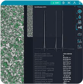
The CellReporterXpress update consisted of new hardware and software autofocus features. The hardware autofocus improvements involve reflectivity detection, during which the autofocus LED is used to find two reflection points at the air-plastic bottom interface and the plastic-liquid interface. The CellReporterXpress-specific algorithms then utilize these two points to find the exact interaction point for proper focus.


Detect surface algorithms

Plate Bottom

Plate Bottom

Hardware autofocus detects the surface closest to the objective (that is, the plate bottom). It works well for adherent samples in plates or chamber slides when imaging at low magnification or if your labware has a thick bottom. This is the fastest hardware autofocus option.

Well Bottom

Well Bottom

Hardware autofocus detects the two surfaces closest to the objective (that is, the plate bottom and well bottom). The Well Bottom option is designed for samples in a liquid medium, such as well plates or chamber slides. This is the most commonly used hardware autofocus option.

Well Insert

Well Insert

Hardware autofocus detects the three surfaces closest to the objective (that is, the plate bottom, well bottom, and well insert). The Well Insert option is designed for well inserts in well plates or any labware design that has a distinct third surface.


In some cases, the highest-contrast imaging focal plane differed from the bottom of the plate. To account for this, Molecular Devices introduced software autofocus routines with varying search ranges to CellReporterXpress, which could find the highest-contrast imaging focal plane automatically.

Learn more about unique and innovative autofocus modes like the 3 Peak Well Insert mode. As well as focus modes designed to enhance acquisition speed, Anchor Focus Position, when imaging samples at low magnification for screening or for large macroscopic samples.


 From widefield to digital confocal

One of the main goals at Molecular Devices is to provide cutting-edge imaging solutions for a broad spectrum of researchers. The ImageXpress Pico system is one of our products that the word affordability has been strongly emphasized. In the continuum of imaging products within the Molecular Devices portfolio, speed, flexibility, and resolution, are the main distinguishing features as you move from the ImageXpress Pico system to the flagship ImageXpress® Confocal HT.ai High-Content Imaging System.
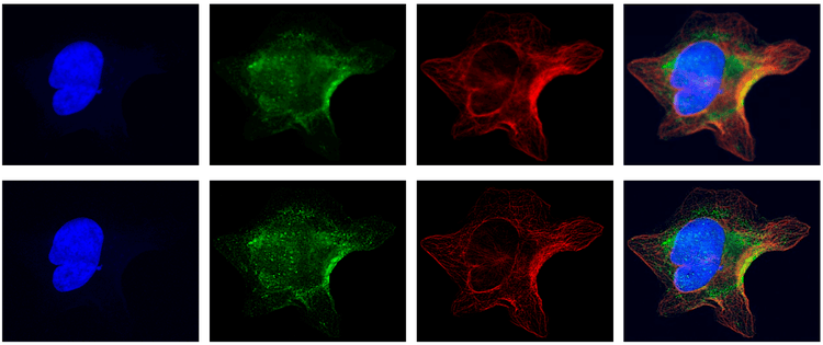
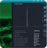
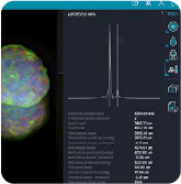
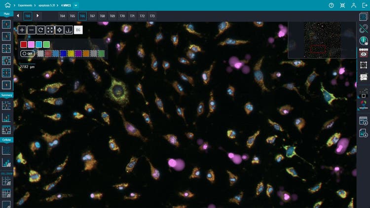
Digital Confocal Option enables robust image analysis

Images with Digital Confocal Option enabled during acquisition (bottom) show cell structures more clearly than without (top), enabling more robust image analysis.

In the case of resolution, we introduced software updates that helped generate confocal-like images from widefield microscopy acquisition. We introduced a restorative image deconvolution method, digital confocal, that quantitatively reassigns out-of-focus light to its original point of origin. Utilizing the point spread function calculated from the specific optical path and objective, the microscopist can designate individual acquisitions to take advantage of increased resolution. Another significant benefit is reduced acquisition time, which meant that phototoxicity risk would also be reduced. Last, but not least, signal-to-noise is enhanced enabling much easier quantification of the images.


 Live preview

Another difference of automated microscopes, in general, was that they did not offer the traditional freedom of movement associated with standard microscope workflows. This created a different user experience, especially in slide-based workflows and cell migration imaging.

The CellReporterXpress software’s new Live Preview feature was developed to tackle this challenge. With Live Preview, using the same camera utilized for acquiring images, the user can now move the sample stage to explore a continually updated, dynamic image of the sample with a virtual joystick, mimicking the traditional experience.

CellReporterXpress software’s Live Preview

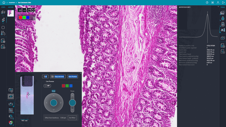
The feature boasts two virtual joysticks that you can use to move around and determine your region of interest at low magnification before image acquisition. The snap overview add-on uses a planetary overview camera to take a snapshot of the entire slide. That way, the user can easily locate their sample on the slide. Once you find the region of interest, the joystick will then help you to seamlessly switch to high magnification for adjusting focus.


Environmental control

Clinically relevant disease models are becoming increasingly significant for mimicking in vivo conditions. This created a shift from endpoint and fixed cell assays to fluorescent or label-free live-cell assays; however, this required thorough control and monitoring of the microclimate around the plates, which is particularly challenging in many imaging systems.

Environmental controls on the ImageXpress Pico system

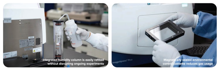
The Environmental Control features in the ImageXpress Pico system provide complete control of the climate, including CO2, humidity, temperature, and O2 (which is extremely useful in modeling hypoxia-based studies, such as those involving tumoroids). Furthermore, the live recording of environmental control sensors enabled users to verify that their cells are being maintained in the ideal microclimate.

The EC settings also enable kinetic acquisitions with multiple time points for different time lengths. The user can generate movies, images, and kinetic data across time, all of which are suitable for export.


Preconfigured protocols with on-the-fly analysis of multiparametric data

Save time and reduce errors with preconfigured analysis protocols that generate robust multiparametric data for a majority of the most common cell-based assays with CellReporterXpress, enabling on-the-fly analysis

Microplate readers have long proven efficient in drug screening and toxicity analysis; however, they are also limited in the information they give, which is fluorescence unit per well. When customers also wanted to track morphology changes and cell behavior in disease models, imaging modalities became critical to compliment the well based data provided by traditional microplate readers.

CellReporterXpress is preinstalled with over 25 preconfigured analysis protocols ranging from simple cell counting to sophisticated multi wavelength cell scoring, and neurite tracing analysis. With features like the click-to-find tool, analysis parameters can be optimized by simply clicking on a few cells that match specific criteria. Allowing for the use of high content analysis techniques to stratify cell populations.

Preconfigured Protocols with Multiparametric Data

For more complex analyses, images captured with the ImageXpress Pico system are compatible with our other Molecular Devices advanced analysis software. The updates in IN Carta® Image Analysis Software and MetaXpress® High-Content Imaging Acquisition and Analysis Software aimed to ease the stratification process. This created a more informed scoring system based on multiple markers within the cell. As for IN Carta software, the SINAP module made use of deep learning algorithms to improve accuracy and flexibility.

In both updates, the user gained a better ability to profile and cluster cell groups, acquire and interpret images, and export & share the resulting graphs.


Lab Automation for high-content screening

Through this paradigm shift to automated imaging, researchers now had access to increased screening throughput utilizing microscopy. The accelerated imaging process naturally necessitated the need for including laboratory automation with the flexibility for novel experimental setups. While some customers wanted to drastically increase their sample size for higher statistical confidence, others wanted to investigate varied disease models with multiple cell lines with a single workflow. The plethora of workflow demands meant that Molecular Devices needed to create customized workflows for various research needs.

Automated ImageXpress Pico workflow


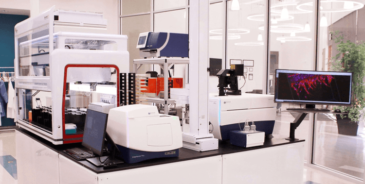
See the Automated ImageXpress Pico system workflow in action

That’s where the automation and customization team came to the rescue. The team provides streamlined workflows that will meet your specific research requirements in the most time-efficient way. The team can modify Molecular Devices’ microplate readers and high-content imaging systems, as well as pair these instruments with automated incubators and liquid handling into seamless workflows. Their workflow customization services involve a wide range of hardware and software modifications, such as the following:

  • Robotics - so you can move plates in and out of the imaging platform with ease
  • Installation of liquid handling workstations
  • Automated reagent addition
  • Automated incubator setup
  • Validation testing

The best part of our automation and customization services is the in-house expertise in instrument and workflow design and continuous support during the consultation, feasibility testing, and implementation.


Organoid Innovation Center: Where motivation meets expertise

Organoid Innovation Center

The relation between Molecular Devices and its customers is bi-directional. As our innovative approach continues to launch products and solutions, our customers continue to thrive in accelerating their research. This drives them to explore more and us to work harder to meet more challenging research questions.

Our new Organoid Innovation Center constitutes a perfect simulation of the compelling customer-team-product relations. To meet research needs for complex 3D biology, we are constantly on the move to keep our imaging and analysis tools up to date. Furthermore, our in-house scientists are available to collaborate with you and provide uninterrupted guidance.

If you are interested in creating an automated workflow for your organoid research, visit our Organoid Innovation Center to learn more about our products and services.

  • Share on LinkedIn
  • Share on Facebook
  • Share on Twitter
  • Share on Youtube

Recent posts

View All Posts
Spacer

How 3D Cell Models Will Shape the Future of Drug Discovery

Target discovery and drug development rely heavily on 2D cell and animal…

A roundtable discussion
Spacer

A case study for assay-ready patient-derived organoids (PDOs) and high-throughput 3D imaging to advance drug discovery

Introduction – the problem.  The average cost of bringing a new drug to…

Read case study
Spacer

3D organoids and automation of complex cell assays [Podcast]

As we enter the era of sophisticated drug discovery with gene therapy and…

Discover 3D organoids
Spacer

Celebrating the art of science: A D+I interview with Daniele Tortorella

At Molecular Devices, we cultivate a culture where associates around the…

Celebrate D+I
Spacer

Advanced technology for automated 3D biology workflows #SLASEurope2022

SLAS Europe 2022, hosted numerous sessions packed with the latest…

View SLAS EU activities
Compare /

Microplate Reader

  • SpectraMax i3x
  • SpectraMax iD3 / iD5
  • SpectraMax M 시리즈
  • SoftMax Pro 소프트웨어

세포 이미징

  • ImageXpress Confocal HT.ai
  • ImageXpress Pico
  • IN Carta 이미지 분석
    소프트웨어

클론 스크리닝

  • ClonePix 2 포유동물 콜로니 피커
  • QPix 미생물 콜로니 피커

FLIPR System

AXON PATCH-CLAMP

실험실 자동화

GxP 컴플라이언스

  • 뉴스
  • 이벤트
2023년 4월 2일

Focus on Microscopy (FOM)

2023년 5월 9일

Imaging User Meeting 2023

2023년 6월 26일

MPS World Summit

행사 더 보기

2023년 2월 22일

Silver Sponsor Molecular Devices at Society for Laboratory Automation and Screening 2023 International Conference and Exhibition

2023년 2월 13일

Molecular Devices inks deal with HUB Organoids to advance automated intestinal organoid screening technology

2023년 2월 7일

Molecular Devices partners with SEED Biosciences to exclusively offer DispenCell Single-Cell Dispenser, expanding leadership in cell line development

뉴스 더 보기
뉴스레터 로더
  • 2023년 3월  
  • 2023년 2월  
  • 2023년 1월  

글로벌 본사

  • 주소 아이콘

    Molecular Devices, LLC.
    3860 N First Street
    San Jose, CA 95134

  • 연락처

    Phone: +1 800-635-5577

    Phone: +86-400-821-3787

    Phone: +44-118-944-8000

    Phone: +44-118-944-8000

    전화: +82-2-3471-9531

    Teléfono: +44-118-944-8000

    Telefono: +44-118-944-8000

  • 사무실 영업 시간

    Mon-Fri 8:00 am - 5:00 pm (PST)

    周一至周五,早上 9 点至下午 5 点 30 分

    Montag bis Freitag 8 h à 17 h (GMT)

    lundi à vendredi 8 h à 17 h (GMT)

    월~금, 오전 9시~오후 5시(한국표준시)

    de lunes a viernes 8 h à 17 h (GMT)

    lunedi al venerdì 8 h à 17 h (GMT)

  • 제품 영업

    Sales +1 877-589-2214

    售前咨询 : 400-821-3787 转 1

    Verkauf 00800 665 32860

    Ventes 00800 665 32860

    판매 +82-2-3471-9531

    Ventas 00800 665 32860

    Saldi 00800 665 32860

  • 기술 지원

    Support +1 800 635 5577

    售后咨询 : 400-821-3787 转 2

    Unterstützung +44-118-944-8000

    Support +44-118-944-8000

    지원 +82-2-3471-9531

    Apoyo +44-118-944-8000

    Supporto +44-118-944-8000

  • 자료 창고

    SpectraNet 고객 포털

바닥글 탐색

  • 이용약관
  • 개인정보 보호정책
  • 데이터를 판매하거나 공유하지 마십시오.
  • 온라인 이용약관
  • 상표
  • 지원
  • 연락처
  • 사이트맵
  • Danaher Life Sciences
  • 채용

©2023 Molecular Devices, LLC. 모든 권리 보유.

©2023 Molecular Devices, LLC. 美谷分子仪器(上海)有限公司保留所有权利。
All Rights Reserved 沪ICP备05056171号-1

barcode