CellReporterXpress
이미지 획득 및 분석 소프트웨어
ImageXpress Pico 시스템을 사용하는 자동화 현미경에 최적화된 익히기 쉬운 소프트웨어입니다.
Cell Counting부터 복잡한 이미지 분석까지를 위한 자동화 현미경 Imaging 시스템
CellReporterXpress 자동화 이미지 획득 및 분석 소프트웨어는 ImageXpress® Pico 시스템에서 작동합니다. 이 시스템은 자동화 현미경으로 획득한 이미지에 대한 정량화된 분석을 수행하기 위한 간단하고 배우기 쉬운 인터페이스를 포함합니다. 이 소프트웨어는 다양한 기능의 이미지의 분석을 통해 처리량을 높힐 수 있으며, 슬라이드 또는 Microplate에 담은 세포 디지털 현미경 이미징의 스케일 조절에 이상적입니다. 다양한 프로토콜과 아이콘으로 표시된 간소화된 워크플로우는 손쉬운 사용자 경험을 제공합니다. 맞춤형 생체모사칩 플레이트를 포함한 다양한 응용 분야를 위한 자동화 초점 알고리즘, Digital Confocal 2D On-the-fly 이미지 데콘볼루션 및 관심 대상 특정 부위 식별을 위한 라이브 미리보기 등의 기능으로, 시간을 절약하는 동시에 assay 품질을 개선하고 해상도를 높일 수 있습니다.

획득 중 분석
당사의 통합된 On-the-fly 이미지 분석 기능으로 데이터 획득 중에도 수치 데이터를 볼 수 있기 때문에 실험을 실행하는 시간을 줄일 수 있습니다. 또한 당사의 디지털 공초점 옵션은 실시간으로 2D 이미지 데콘볼루션을 수행하므로, 해상도와 assay 품질을 상당히 높여 줍니다.

이미지 분석 간소화
Label-free 및 형광 이미지용 분석 프로토콜을 사용하면 일반적인 생물학적 assay를 신속하게 실행하는 기능을 제공 받는 동시에 자체 프로토콜을 생성 및 저장할 수 있습니다. 단순한 Cell Counting에서 복잡한 Neurite Tracing에 이르기까지, 그리고 2 및 3레인 Mimetas 프로토콜이 포함된 다양한 최적의 템플릿을 사용하면, 사용자가 더 이상 복잡한 Parameter를 조정하거나 확실한 분석을 위해 제3자 소프트웨어로 내보내는 것에 대해 우려할 필요가 없습니다. IN Carta® 이미지 분석 소프트웨어를 추가하면 기계 학습을 활용하여 더 많은 정보를 얻고 데이터 분석의 정확도를 높여 자신 있게 새로운 발견을 가능하게 할 수 있습니다.

어디서나 데이터를 공유하고 액세스하세요.
이 소프트웨어를 사용하면 동료와의 공유, 프레젠테이션 및 협업이 더 쉬워지며, 여러 사용자가 브라우저 기반 원격 액세스를 통해 소프트웨어를 동시에 사용할 수 있습니다.
https://view.ceros.com/molecular-devices/imagexpress-pico-digital-confocal/p/1
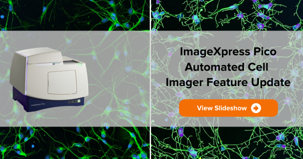
CellReporterXpress의 새로운 소식 보기
기능

25개 이상 내장된 응용 분야 프로토콜
사용자 지정 인공 지능 루틴은 Parameter를 자동으로 최적화하여 견고하고 사용하기 쉬운 분석 프로토콜을 생성합니다.

다양한 세포 기반 Imaging 응용 분야를 위한 자동 초점 루틴
다양한 샘플 유형에 대한 고품질 Imaging을 보장하기 위해 모든 실험 기구에 안정적인 초점을 제공합니다.

이미징 획득 부위 선택
핵심 관심 대상 영역만을 획득 및 분석하기 위해 Well 또는 슬라이드 내 다양한 부분들을 선택할 수 있습니다.

Label-Free 분석
Brightfield에서 소형 원형 세포, 대형 세포 및 Bead 등의 대물을 자동으로 식별합니다.


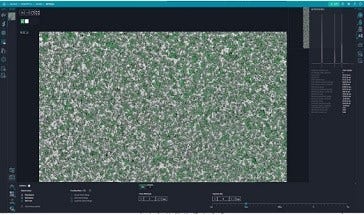
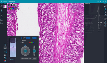
Side by Side 세포 확대
전체 Well 보기부터 세포 수준 보기까지 고해상도 이미지를 확대/축소 또는 패닝합니다. 두 Well의 이미지를 동시에 비교합니다.

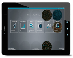
터치스크린 기능
탁월한 사용성 및 정밀한 제어 기능이 개인용 컴퓨터 또는 태블릿 사용에 관계없이 내장되어 있습니다.

브라우저 기반 소프트웨어
ImageXpress Pico 시스템은 사용자 네트워크에서 모든 컴퓨터로부터 받은 데이터를 Chrome™ 또는 Safari®을 통해 브라우저에서 제어할 수 있습니다.
지능형 이미지 획득 및 분석
ImageXpress Pico 시스템과 함께 CellReporterXpress 소프트웨어는 Imaging 이상의 효과를 제공합니다. Cell Based Assay를 위한 이미지 분석을 간소화하는 최고의 분석 기능을 제공합니다.
자동 초점
다양한 응용 분야를 위한 자동 초점 알고리즘
두 개의 견고한 자동 초점 메커니즘: 하드웨어 자동 초점을 위한 Detect Surface 및 이미지 기반 자동 초점을 위한 Find Best Plane. 다양한 샘플 유형에 대한 고품질 Imaging을 보장하기 위해 모든 실험 기구에 안정적인 초점을 제공합니다.
디지털 공초점(Digital Confocal)
on-the-fly deconvolution으로 해상도 증가
Digital Confocal* 2D On-the-fly 데콘볼루션 옵션으로 획득하는 도중 이미지의 대비를 향상시켜 해상도를 높이고 분석 품질을 개선할 수 있습니다.
Live Pre-view
관심 영역(ROI)을 신속하고 손쉽게 식별하기
라이브 미리보기 기능을 사용하면 가상 조이스틱을 이용하여 샘플의 초점을 조정할 수 있으므로 관심영역(ROI)을 확인하는 과정이 간편해지고, 빠르고 손쉽게 작업할 수 있습니다.
EC(Environmental Control)

Environmental Control System으로 Live Cell Assay를 Monitoring
장기간 동안 수행되는 Time lapse 및 Live Cell assay는 습도, CO2 및 O2 제어를 위한 옵션이 포함된 Environmental Control System을 사용하여 실행할 수 있습니다. 또한 z 표동을 방지하기 위해 최적화된 이 소프트웨어는 환경 상태를 실시간으로 모니터링하여, 최적의 assay 조건을 보장합니다.
다중 파장 세포 점수화(Cell Scoring)
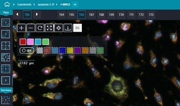
최대 4개의 형광 염색으로 다중 파장 세포 점수화 기능
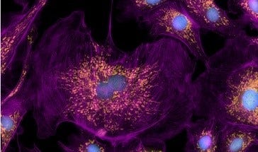
내장된 프로토콜은 여러 파장 실험에서 세포의 counting 및 기록 측정에 이상적입니다. 핵에 대한 형광 표지자와 세포질에 대한 추가 표지자를 사용하여 각 파장을 분석하고 세포에 다중 매개변수 표현형 프로파일을 할당합니다.
Z-Stack 획득
Z-stack 획득으로 더 깊은 통찰력 확보
z-stack 획득을 사용하여 좀 더 정확한 세분화를 위한 더욱 선명한 이미지를 생성할 수 있습니다. 서로 다른 초점에서 일련의 이미지를 획득하여 단일 절편(Slice)보다 좀 더 세부적인 정보를 얻을 수 있습니다. 사용자들은 모든 절편을 포함시키거나 최종 이미지에 어떤 절편을 포함시킬 것인지 선택할 수 있습니다.
내장 분석 프로토콜로 시간 절약 및 오류 감소
단순한 Cell Counting부터 정교한 Neurite Tracing 분석에 이르기까지 다양한 25개 이상의 내장 분석 프로토콜이 있습니다. 클릭 기능 검색 도구와 같은 기능을 통해, 특정 기준에 맞는 몇 개의 세포를 클릭하기만 해도 분석 Parameter를 최적화할 수 있습니다.
각 CellReporterXpress 기능에 대한 자세한 내용은 아래 아이콘 클릭
CellReporterXpress는 ImageXpress Pico Automated Cell Imaging System의 일부입니다. 자세히 알아보려면 ImageXpress Pico 제품 페이지를 방문해 주십시오.
자동 Brightfield, 형광 및 Digital Confocal 이미징의 디지털 현미경
- Cell Counting, Transfection Efficiency 및 cell health assay에 적합
- 하드웨어 자동 초점은 빠른 속도를 위해 LED 빔을 사용하여 반사 표면을 찾기 위해 설계되었습니다.
- Environmental Control System으로 Live Cell Assay를 Monitoring
- 브라우저 기반, 태블릿 및 터치스크린 호환 소프트웨어
- 로보틱스, 배양기 및 소프트웨어를 이용한 맞춤형 실험실 자동화 솔루션
최신 자료
당신의 다음 연구와 새로운 발견을 위해 어떤 도움이 필요하신가요?
뛰어난 역량의 직원들이 고객분들의 연구과제의 문제 해결을 위해 일선에서 대기하고 있습니다.
원격 또는 현장 제품 데모, 웨비나 등을 통해 도움을 받으실 수 있습니다. 무엇을 도와 드릴까요?
아래 항목 중에서 선택하십시오.