신경 발달 및 퇴행의 𝘪𝘯 𝘷𝘪𝘵𝘳𝘰 연구를 위한 Neurite Outgrowth assay를 이용한 뉴런의 특성 분석 간소화
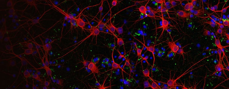
뉴런은 축색돌기과 수상돌기로 불리는 세포체의 확장을 통해 연결을 만들어내며, 이를 흔히 ‘신경돌기’ 또는 ‘돌기’라고 부릅니다. 이러한 생물학적 현상을 Neurite Outgrowth라고 하며, 이는 복잡한 세포 내 신호전달 작용에 의해 조절됩니다.
Neurite Outgrowth는 생체 외에서 신경 발달 및 신경 퇴행을 연구하기 위해 일반적으로 사용되는 assay입니다. 신경돌기의 발달에는 세포 외 및 세포 내 신호 모두의 복잡한 상호 작용이 필요합니다. 신경돌기의 생장은 신경 영양 인자에 의해 자극 또는 억제될 수 있습니다. 중요한 것은, 뉴런의 발달이 신경독성 화학물질에 의해 영향을 받을 수 있다는 점입니다.
Neurite Outgrowth을 촉진하는 신호전달 기전을 이해하게 되면, 신경독성 반응, 화합물 스크리닝 데이터, 신경 발달과 재생 분석에 영향을 미치는 요인과 관련된 가치 있는 통찰을 얻을 수 있습니다. Neurite Outgrowth의 억제나 자극은 뇌졸중, 파킨슨병, 알츠하이머병, 척수 손상을 포함한 광범위한 CNS 장애 또는 손상을 의미합니다.
Neurite Outgrowth 분석을 위한 실험과정 솔루션
Neurite Outgrowth는 신경 단위 프로세스의 세분화 및 정량화를 통해 평가됩니다. 이러한 신경 돌기는 형광 현미경을 이용하여 이미징할 수 있으며, 처리량이 적은 경우에는 수동 추적과 계수로 정량화할 수 있습니다. 그러나, 더 처리량이 많은 Microplate 형식의 샘플의 경우에는 자동화 Imaging 시스템을 분석 소프트웨어와 함께 사용하는 것이 더 효율적인 솔루션입니다.
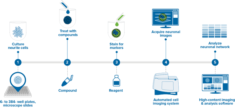
이 실험과정은 간소화된 Neurite Outgrowth 분석 과정을 설명하며, 효율적인 연구와 처리량 증가를 돕는 시스템을 중점적으로 살펴봅니다.
- 신경 세포 배양 – 세포를 배양한 뒤 96 또는 384 Well Microplate에서 신경돌기망을 형성하도록 하였습니다.
- 화합물 처리 – 이후, 세포는 48시간 동안 독성 화합물에 노출되었습니다.
- 표지용 염색– 화합물 처리가 완료된 뒤, Live Cell 염색액을 배지에 직접 첨가할 수 있습니다. 형광 접합 항체를 이용한 면역염색법 프로토콜은 세포 고정 이후에도 수행할 수 있습니다.
- 뉴런 이미지 획득 – 뉴런의 High-Content Imaging으로 과학자는 신경돌기의 수, 길이, 분지화와 같은 신경 네트워크의 변화 특성 분석과 측정을 비롯해 육안 또는 특정 독성 반응을 모두 파악할 수 있게 되었습니다. 넓은 시야의 이미지를 획득하여 웰당 더 적은 부분에서 더 많은 세포를 채취할 수 있으므로 플레이트 획득 시간이 크게 단축됩니다.
- 뉴런 네트워크 분석 – High Content Analysis는 Neurite Outgrowth 분석에 대한 긍정적, 부정적 요인의 영향을 파악하는 정량적인 방법을 제공합니다. 세포 이미징 분석 소프트웨어를 이용한 세포당 돌기 수, Neurite Outgrowth 길이, 분지화, 세포의 수를 포함한 여러 Parameter의 특성 분석을 위해 신경세포 이미지를 정량적으로 분석하십시오.
Neurite Outgrowth의 응용 분야 및 assay
뉴런의 High-content imaging으로 과학자들은 신경돌기의 수, 길이, 분지화와 같은 신경 네트워크의 변화 특성 분석과 측정을 비롯해 육안 또는 특정 독성 반응을 모두 파악할 수 있게 되었습니다.
자동화 현미경과 high-content analysis 소프트웨어를 이용하여 신속하고 정확하게 신경 활성을 캡처하고 정량화하는 방법에 대해 알아보십시오.