암 연구 솔루션
암 연구에서 cellular pathway와 세포 간 상호작용의 이해를 위한 생명현상 분석 솔루션
암 치료 스크리닝을 위한 spheroid, 오가노이드, organ-on-a-chip 생물학과 같은 세포 모델의 3D 이미징과 분석
암은 세포가 정상 한계치 이상으로 생장 및 분열하고, 인접 조직을 칩입 및 파괴하며, 궁극적으로는 체내의 원발 부위로 전이하는 변화와 관련이 있습니다. 암 연구자에게는 복잡하거나 잘 알려지지 않은 암세포와 세포 환경 간의 상호작용을 보다 용이하게 연구하고 치료적 중재 지점을 식별하기 위한 도구가 필요합니다.
종양과 장기의 in vivo 환경을 더욱 닮은 스페로이드, 오가노이드, 생체모사칩 시스템과 같은 생물학적 연관성을 가진 3D 세포 모델을 이용한 암 연구가 가능하도록 하는 우리의 High-Content Imaging 시스템과 High-Content Analysis 소프트웨어 솔루션에 대해 알아보세요.
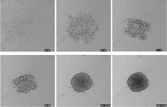
그림 1: High-Throughput Screening 환경에서 스페로이드 테스트를 위한 실험과정 단일 스페로이드를 96 또는 384 well 플레이트에서 성장시키고 약물로 처리하고 세척 없이 imaging할 수 있는 혼합 염료시약으로 염색할 수 있습니다. 원하는 경우 스페로이드도 고정할 수 있습니다. (우측) ImageXpress Micro confocal 시스템에서 Timelapse 획득을 사용하여 63시간 동안 HCT116 세포의 투과광 이미지를 촬영하여 스페로이드 형성을 보였습니다(10X 대물렌즈).
Cancer Spheroid에 있어 3D 이미징 기술의 장점
암 스페로이드는 기존 2D 세포 배양에 비해 종양 행동을 훨씬 효과적으로 모방합니다. 이 3D Spheroid 모델은 가능성 있는 암 치료를 식별하는 환경 스크리닝에 성공적으로 이용되고 있습니다. 이 배양 시스템은 다양한 생명현상의 결과물을 정량화하기 위한 다중 파라미터 분석에 이용되어 암 치료제 개발을 가속화시킬 수 있습니다.
주요 이점:
- 3D high-content imaging의 개발은 정확도와 관련성이 더 높은 검사를 쉽게 가능하도록 하는 주요 단계로서 의미가 있습니다.
- 3D 배양 시스템은 High-throughput 포맷에서 사용할 수 있는 균일한 인간 cancer cell spheroid를 빠르게 생산하여 암 치료제 개발을 가속화할 수 있습니다.
- 암세포를 대상으로 한 confocal 3D image 분석을 이용한 연구는 여러 생명현상의 결과물에 대한 다중 파라미터 특성을 분석할 수 있습니다.
High-Throughput Screening 환경에서의 3D Cancer Spheroid 분석 실험과정
스페로이드는 96 또는 384 well 플레이트에서 성장시키고 약물로 처리하고 현장에서 세포의 작용 과정과 경로를 보여주는 염색시약으로 염색할 수 있습니다. 몇몇 경우에는 스페로이드를 세척 없이도 이미징할 수 있으며, 필요에 따라 고정할 수도 있습니다.
다양한 이미징, 세포 스크리닝, Microplate Reader 시스템으로 Oncology 연구의 실험과정을 간소화하십시오.
워크플로는 간소화된 스페로이드의 분석 절차를 보여주며, 연구를 효율적으로 간략화하고 처리량을 높일 수 있는 시스템을 강조합니다.
스페로이드 배양 – 암세포는 ULA(ultra-low attachment), 둥근 바닥 플레이트 또는 다른 실험기구에 직접 배양하여 일반적인 스페로이드 형태로 발생하도록 할 수 있습니다. 다른 실험기구로 암세포를 단일 웰에서 다중 스페로이드로 배양할 수 있습니다.
화합물 처리 – 스페로이드가 형성된 이후에 원하는 농도의 화합물을 Well에 첨가한 뒤 연구 중인 기전에 따라 하루에서 수 일 동안 배양합니다.
표지자 염색하기 – 화합물 처리가 완료되면 염색액을 배지에 직접 첨가합니다. 스페로이드가 교란되지 않도록 세척이 필요하지 않은 염색액을 사용할 수도 있지만, 필요하다면 자동화 기기를 사용해서라도 스페로이드를 조심스럽게 세척해도 됩니다.
스페로이드 이미지 획득하기 – 스페로이드의 체내 이미지는 개별적으로 캡처하거나, 특수 이미징 기기를 이용하여 Z-stack(여러 깊이에서 촬영한 다중 이미지)으로 캡처할 수 있습니다.
암 세포 분석하기 – 세포 이미징 분석 소프트웨어의 세포 이미지에 대한 정량적 분석을 실행하여 다양한 표지자의 발현을 Monitoring하고 생물학적 판독값을 정량화합니다.
암 치료의 스크리닝을 위한 spheroid의 High-Throughput Confocal Imaging
최근 수년간, in vivo 조직 환경 모델로 사용되는 in vitro 종양 세포 Aggregate 형성에 상당한 진전이 있었습니다. 저부착성 둥근 바닥 Microplate의 Well에 시드(seed)하는 경우, 이 세포 응집체는 별개의 spheroid를 형성합니다. 스페로이드는 양 표면이 노출되고 깊이 분포한 세포, 생장 및 비생장 세포, 세포의 저산소성 중심부와 산소 공급이 잘 되는 세포 외층을 포함하여 종양과 매우 유사하므로, 기존의 2차원 배양 세포보다 종양 행동을 더 잘 모방하는 것으로 여겨집니다. 이 3D Spheroid 모델은 가능성 있는 암 치료를 식별하는 환경 스크리닝에 성공적으로 이용되고 있습니다.
확실한 spheroid assay의 개발에는 여러 어려움이 있지만, 자동화 High-throughput을 이용한 high-content imaging은 항암화학 요법 후보 약물과 관련된 검사의 연관성을 높일 수 있는 유의미한 단계입니다.
- 20X 배율의 단일 관측 시야로 전체 스페로이드를 캡처
- 96 또는 384 well 형식에서 생물학적으로 연관성을 가진 3D 스페로이드 Screening
- 정확한 세포 반응 감지를 위해 confocal imaging을 사용
- Z 평면 이미지의 2D 재구성만 저장하여 저장 공간을 절약
그림 1. 마이크로플레이트의 3D 스페로이드의 신속 스크리닝
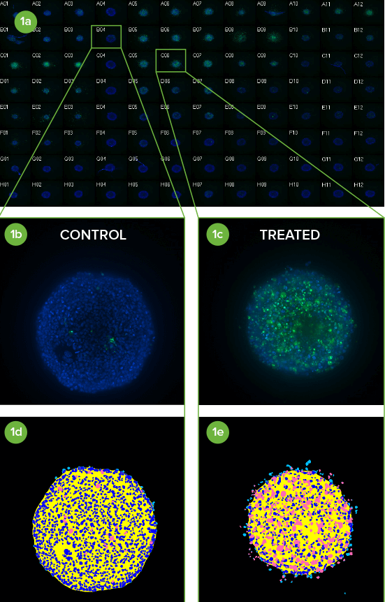
(1a) 10X Plan Flour Objective(대물렌즈)를 사용하여 화합물 처리한 96 Well Plate의 HCT116 Spheroid의 이미지 섬네일 몽타주. Hoechst 염색된 핵(청색)에 CellEvent® Caspase3/7세포사멸 표지자(녹색)를 중첩하였습니다.
(1b) 컬럼 내의 미처리 대조군4과 (1c) A열에 Paclitaxel을 1µM부터 1:3로 연속 희석한 5–7 컬럼 내 Caspase 3/7 반응이 분명하게 보입니다(3의 반복 실험).
(1d, 1e) 11개의 z 평면을 2D Maximum Projection 이미지로 통합하고 간단한 맞춤 모듈로 분석하였습니다. 낮은 세포사멸 정도와 높은 세포사멸 정도를 이에 상응하는 분할 마스크(감청색 = 핵, 분홍색 = 사멸세포)로 나타낸 원본 이미지.
응용 분야 및 Assay
Molecular Devices는 세포 이미징의 업계 선두주자로서, 생명과학 연구, 신약 개발, high-throughput screening을 지원하는 다양한 도구를 제공합니다. 당사의 high-content imaging 시스템은 생명현상 분석을 이용한 암 연구 활동을 성공으로 이끌 수 있습니다. 또한 당사는 Multi-Mode Microplate Reader와 더불어 사용이 편리한 마이크로어레이 스캐너에 대한 여러 가지 구성을 제공합니다.
당사의 기술이 암 치료 연구를 어떻게 도울 수 있는지 더 살펴보십시오.