Molecular Devices, LLC
  •   美谷分子仪器(上海)有限公司
    咨询服务热线:400-821-3787
  •   0 Cart
  • Store

  •  채용
  •  언어  
    • English
    • Deutsch
    • 中文
    • 日本語
    • Français
    • 한국어
    • Italiano
    • Español
Molecular Devices, LLC
  • 제품
    • 제품

      새로운 DispenCell™ Single-Cell Dispenser 기술은 단일 세포주를 3배 더 빠르고 저렴한 비용으로 분리합니다.

      • 쉽고 직관적인 설정
      • 클론형성능과 추적 가능성의 즉각적인 증거
      • 고유한 기술로 세포 샘플을 부드럽게 처리
      • 벤치탑 크기  
        설계
      • 특허받은  
        일회용 팁
      DispenCell™ Single-Cell Dispenser 기술

      최적화된 3D 조직과 오가노이드 실험과정을 위한 BioAssemblyBot의 6축 로봇 팔을 갖춘 자동화된 High-Content Screening 솔루션

      BioAssemblyBot의 6축 로봇 팔

    • Microplate Reader
      SpectraMax Mini Multi-Mode Microplate Reader
      Multi-Mode Reader
      • SpectraMax i3x
      • SpectraMax iD3/iD5
      • SpectraMax M 시리즈
      • FlexStation 3
      • SpectraMax Mini
      SpectraMax ABS 마이크로플레이트
      흡광(Absorbance) 리더기
      • SpectraMax ABS/ABS Plus
      • SpectraMax VersaMax
      • SpectraMax QuickDrop
      • CMax Plus
      Fluorescence Reader
      Fluorescence Reader
      • SpectraMax Gemini
      SpectraMax Luminescence
      발광(Luminescence) 리더기
      • SpectraMax L


      MultiWash+ Washer
      Stacker 및 Washer
      • StakMax Stacker
      • AquaMax Washer
      • MultiWash+ Washer
      • MultiWash–C 微孔板洗板机
      SoftMax Pro 데이터 획득
      분석 소프트웨어
      • SoftMax Pro 소프트웨어
      • SoftMax Pro GxP Software
      GxP 솔루션
      GxP Compliance Solution
      • SoftMax Pro GxP Software
      • 소프트웨어 설치와 Validation 서비스
      • IQ/OQ/PM 서비스
      • SpectraTest Validation Plate
      실험실 자동화와 맞춤화
      실험실 자동화와 맞춤화
      • 플레이트 기반 High-Throughput Assay를 위한 실험실 자동화
    • 세포 Imaging 시스템
      ImageXpress Pico Automated Cell Imaging System
      Automated Cell Imaging Systems
      • ImageXpress Pico
      • ImageXpress Nano
      High-Content Imaging
      High-Content Imaging
      • ImageXpress Confocal HT.ai
      • ImageXpress Micro Confocal
      • ImageXpress Micro 4
      StratoMineR 분석
      Acquisition & Analysis Software
      • IN Carta
      • StratoMineR
      • MetaXpress
      • CellReporterXpress
      • MetaMorph
      실험실 자동화와 맞춤화
      실험실 자동화와 맞춤화
      • High-Throughput, High-Content Screening(HCS)을 위한 실험실 자동화
      • BioAssemblyBot 400 Bioprinter 자동화 HCS 솔루션
    • 클론 스크리닝
      Clone Pix 시리즈
      포유류 콜로니 피킹
      • ClonePix 2
      QPix 미생물 콜로니 피커
      미생물 콜로니 피킹
      • QPix 420
      • QPix 450/460
      • QPix HT
      CloneSelect Imager FL
      단일 세포 Imaging
      • CloneSelect Imager
      • CloneSelect Imager FL
      DispenCell Single-Cell Dispenser
      단일 세포 분리
      • DispenCell Single-Cell Dispenser
      실험실 자동화
      실험실 자동화와 맞춤화
      • High-Throughput 클론 스크리닝을 위한 실험실 자동화
      CloneMedia와 XP Media
      배지 및 시약
      Clone Screening Assay kit
      Clone Screening Assay kit
    • FLIPR Penta
      FLIPR Penta
      FLIPR Penta
      • FLIPR Penta High-Throughput 세포 스크리닝 시스템
      Screenworks
      분석 소프트웨어
      • ScreenWorks 소프트웨어
      • Peak Pro 2 소프트웨어 모듈
      Flipr Assay 키트
      FLIPR Assay 키트
      • 칼슘 Assay 키트
      • 칼륨 분석 키트
      • 막전위 분석키트
      • EarlyTox 심장 독성 키트
    • Axon Patch-Clamp
      투명
      Amplifier
      • Axopatch 200B 콘덴서
      • MultiClamp 700B
      • Axoclamp 900A
      투명
      Digitizer
      • Axon Digidata 1550B Low
      투명
      분석 소프트웨어
      • pCLAMP 11 소프트웨어 제품군
    • 기타
      Threshold Immunoassay System
      Threshold Immunoassay System
      Geneppix 미세배열 스캐너
      Genepix 미세배열 스캐너
      Imagexpress Micro Xls
      Imagexpress Micro xls
      인증 리퍼브
      인증 리퍼브
      IDBS 솔루션
      IDBS R&D 클라우드 솔루션
    • 분석 시약(assay kit)
      심장 독성
      • EarlyTox 심장 독성 키트
      Cell viability
      • EarlyTox Cell Integrity Kit
      • EarlyTox Cell Viability Assay Kit
      DNA 정량 분석
      • Spectramax Quant dsDNA Assay 키트
      Elisa, western blot
      • CatchPoint SimpleStep ELISA 키트
      • ScanLater Western Blot Assay 키트
      gpcr
      • FLIPR 칼슘 Assay 키트
      • Fura-2 QBT 칼슘 키트
      • CatchPoint cAMP 형광 Assay 키트
      • CatchPoint cGMP 형광 Assay 키트
      이온 통로
      • FLIPR 칼륨 Assay 키트
      • FLIPR 막전위 Assay 키트
      IGG 정량분석
      • ValitaTiter
      • CloneDetect
      Reporter gene
      • Spectramax Glo Steady-Luc Reporter Assay Kit
      • Spectramax DuoLuc Reporter Assay Kit
      Transporter
      • QBT Fatty Acid Uptake Assay Kit
      • 신경전달물질 수송체 흡수 Assay 키트
      기타
      • Contamination Detection
      • Enzyme - IMAP Assay
    • 액세서리 및 소모품
      Microplate Reader
      • 384 Well SBS
      • 384 Well High Sample Recovery Plate
      • Deep-well Plate
      • Low Profile Microplate
      • SpectraDrop Micro-Volume Microplate
      • SpectraMax Injection cartridge with SmartInject Technology
      • SpectraMax MiniMax 300
        Imaging Cytometer
      • Western Blot Cartridge
      • 96孔微孔板
      클론 스크리닝
      • Adjustable Petri Dish and Microplate Holder
      • Bioassay QTrays
      • Calibead
      • 캡 매트 및 뚜껑
      • 크로마 필터
      • 세정 및 살균 솔루션
      • CloneSelect Single-Cell Printer Cartridge
      • QPix 핀 및 헤드
      • QReps 레플리케이터
      Axon Patch-Clamp
      • Soft Panel Amplifier Control
      Spectra Img
  • 연구 분야
    • 응용 분야

      Molecular Devices, Cellesce 인수로 환자 유래 오가노이드 특허 기술 추가

      2022년 12월 6일

      • Cellesce의 동종 업계 최초 기술로 대규모 약물 Screening을 위한 일관적인 환자 유래 오가노이드를 생성합니다.
      • 인수를 통해 Molecular Devices의 3D 생물학 솔루션 혁신 기업으로서의 입지를 강화합니다.
      • 결합된 전문 지식으로 신약 개발을 위해 생리학적으로 연관된 세포 모델의 채택을 업계에서 가속화할 것입니다.


      OIC 방문하기

      언론 보도 읽기

      Cellsce
      응용 분야를 위한 Spectra
    • 코로나바이러스 (COVID-19)
      코로나19
      코로나19 연구
      관련 솔루션
      코로나19
      코로나19 관련
      새로운 뉴스
      코로나19
      백신 개발 워크플로
      감염병 연구 응용 분야
      백신 연구
      연구 응용 분야
    • 연구분야 (Stem Cell, Cancer)
      투명
      3D Cell Model
      투명
      암 연구 솔루션
      투명
      Cell Line Development
      투명
      신약 개발
      투명
      식품 및 음료
      투명
      유전자 편집(CRISPR/Cas9)
      투명
      오가노이드 연구
      투명
      줄기세포 연구
      투명
      독성학
    • Microplate Reader
      투명
      세포 건강 상태 (Cell Health)
      투명
      Cellular Signaling
      투명
      ELISA
      투명
      미생물학과 오염물질
      투명
      핵산(DNA/RNA) 측정과 분석
      투명
      단백질 측정, 정량분석, 분석
      투명
      관련 분석법: 측정 모드
      • 흡광(Absorbance)
      • 형광(Fluorescence)
      • 형광 편광
      • 발광(Luminescence)
      • TRF, TR-FRET 및 HTRF
      • Western Blot
    • 세포 Imaging 시스템
      투명
      Cell Counting
      투명
      세포 이미징 및 분석
      투명
      세포 Migration Assay
      투명
      세포 염색
      투명
      Live Cell Imaging
      투명
      Neurite Outgrowth
      투명
      장기 칩
      투명
      오가노이드
      투명
      Spheroids
    • 클론 스크리닝
      투명
      Cell line development 실험과정
      투명
      단클론항체(mAb)
      • 하이브리도마
      • 파지 디스플레이
      • 단일클론항체 생산
      투명
      Monoclonality
      투명
      합성 생물학
    • FLIPR Penta
      투명
      GPCR(G protein-coupled receptor)
      투명
      이온 채널
      투명
      Cardiotoxicity(심장독성)
    • Axon Patch-Clamp
      투명
      전기생리학(Patch Clamp)
  • 자료
    • 자료
    • 관련 자료 검색
      메뉴 자료 아이콘/응용 분야 노트
      Application Note
      메뉴 자료 아이콘/레퍼런스
      레퍼런스
      Ebook 아이콘
      eBook
      메뉴 자료 아이콘/Scientific Poster
      Scientific Poster
      메뉴 자료 아이콘/튜토리얼 및 영상
      동영상 및 웨비나

      검색

    • 블로그 – 실험실 노트
      스페이서
      Assay에 대한 고객 사례…
      스페이서
      3D organoids and…
      How 3D Cell Models Will Shape the Future of Drug Discovery
      2023년 3월 7일 Target discovery and drug development rely heavily on 2D cell and animal models to decipher efficacy and toxic effect of drug candidates. Yet, 90% of candidates fail to…
      더 알아보기  
    • 고객 사례 소개

      다른 연구자들이 제품과 솔루션을
      어떻게 사용했는지 확인해보세요.

    • 혁신 기술
      투명
      AI, 머신러닝 및 딥러닝
      투명
      AgileOptix 스피닝 디스크 기술
      투명
      자동 초점
      투명
      디지털 공초점 옵션
      투명
      고함량 Screening
      투명
      HumSilencer
      투명
      레이저 조명
      투명
      QuickID 표적 이미지 획득
    • 관련 분석법
      투명
      흡광(Absorbance)
      투명
      전기생리학
      투명
      형광(Fluorescence)
      투명
      형광 편광(FP)
      투명
      발광(Luminescence)
      투명
      TRF, TR-FRET 및 HTRF
      투명
      Water Immersion Objective
      투명
      Western Blot
    • 동영상 갤러리
      투명
      Microplate Reader
      투명
      세포 Imaging 시스템
      투명
      Flipr 시스템
      투명
      클론 스크리닝
      투명
      Axon Patch-Clamp
      투명
      주문형 웨비나
  • 서비스 및 지원
    • 서비스 및 지원
    • 개요
      Spectra 로고
      SpectraNet 고객 관리 포털
      GxP 컴플라이언스
      GxP 컴플라이언스 솔루션
      실험실 자동화와 맞춤화
      실험실 자동화와 맞춤화
      전문 서비스
      전문 서비스

      기술 지원

      미국 본사  
      +1 800-635-5577  
      월~금, 오전 7시~오후 5시 PST

      유럽  
      +44-118-944-8000  
      월~금, 오전 8시~오후 5시 GMT

      내가 있는 지역의 담당자 확인하기

      SpectraNet 고객 포털  

    • 고객 포털 - Spectranet
      Spectranet

      INTRODUCING OUR NEW CUSTOMERCARE PORTAL

      SpectraNet is an intuitive, simple-to-use, self-service customer portal providing a new level of experience available 24/7.

      Create your account today to get full access to integrated content and world-class customer service.

      REGISTER LOGIN
      지원 티켓 신청
      지원 티켓 신청하기
      자료 창고
      지식 기반
      제품 등록
      제품 등록하기
      기술 자료
      기술 자료
    • GxP 컴플라이언스 솔루션
      GxP Softmax Pro GxP 소프트트웨어
      SOFTMAX PRO GXP 소프트웨어
      GxP 소프트웨어 설치
      소프트웨어 설치와 Validation 서비스
      GxP Spectratestt Validation Plate 재인증
      SPECTRATEST Validation Plate
      IQ OQ 서비스
      IQ/OQ/PM 서비스
    • 실험실 자동화
      실험실 자동화와 맞춤화
      실험실 자동화와 맞춤화
      High Content Screening HCS
      High-Throughput, High-Content Screening
      • BioAssemblyBot 400 Bioprinter 자동화 HCS 솔루션
      플레이트 기반 High-Throughput Assay
      플레이트 기반 High-Throughput Assay
      High-Throughput 클론 스크리닝
      High-Throughput 클론 스크리닝
    • 전문 서비스
      전문 서비스
      전문 서비스
      서비스 플랜
      서비스 플랜
  • 회사
    • 회사

      Molecular Devices, 오스트리아의 글로벌 R&D 허브 확장

      2022년 10월 12일        
      더 큰 부지는 Cell Line Development, 오가노이드 개발, 신약 개발을 개선하기 위한 Screening 솔루션을 발전시키기 위한 협업 공간인 잘츠부르크의 오가노이드 혁신 센터의 미래 기지가 될 것입니다.


      OIC 방문하기

      언론 보도 읽기

      Austrian Research & Development Center의 리본 커팅 기념식
      응용 분야를 위한 Spectra
    • 회사 소개

      Molecular Devices는 실리콘 밸리를 기반으로 제약 및 연구의 스크리닝과 효과적인 분석이 가능한 솔루션을 40년에 걸쳐 개발하고 공급해 왔습니다.

    • 리더십

      당사의 다양한 경험, 비즈니스 인사이트 및 공동의 목표의식은 직원들이 잠재력을 최대한 발휘하도록 독려하기 위한 당사의 일상적인 결정에 추진력을 부여합니다.

      리더십

    • 채용

      당사의 팀 중심 기업 문화는 생각과 관점의 다양성과 강력한 신뢰 관계를 보장합니다.

    • 뉴스룸
      투명
      뉴스
      투명
      보도 내용
      Silver Sponsor Molecular Devices at Society for Laboratory Automation and Screening 2023 International Conference and Exhibition
      Feb 22, 2023 Showcasing new industry collaborations, automated technology, and workflow innovations that span 3D biology, cell line development, and drug…
      Read more  
    • 이벤트
      Focus on Microscopy (FOM)
      Conference | Europe | Porto, Portugal, Europe– Apr 02 – Apr 5, 2023 FOM2023 continues a long-standing (since 1988), yearly conference series on the latest innovations and developments in (optical) microscopy and their…
      Read more  
      Imaging User Meeting 2023
      Conference | Europe | Copenhagen, Denmark– May 09 – May 10, 2023 Ideal for both current ImageXpress system users and those wanting to learn more about high-content, high-throughput, automated or 3D imaging, our…
      Read more  
  • 연락처
  • 검색

    검색

  • 견적
    요청하기

Mobile navigation

  • Products
    • All Products
    • Microplate readers
      • Multi-Mode Readers
      • Absorbance Readers
      • Fluorescence Readers
      • Luminescence Readers
      • Microplate Stacker
      • Microplate Washers
      • SoftMax Pro Software
      • SoftMax Pro GxP Software
      • GxP Compliance Solutions
      • Lab Automation & Customization
    • Cellular Imaging Systems
      • Automated Cell Imaging Systems
      • High-Content Imaging
      • Acquisition & Analysis Software
      • Lab Automation & Customization
    • Clone Screening
      • Mammalian Colony Picking
      • Microbial Colony Picking
      • Single-Cell Imaging
      • Single-cell Isolation
      • Lab Automation & Customization
      • Culture Media & Reagents
      • Clone Screening Assay kit
    • FLIPR Penta
      • FLIPR Penta
      • ANALYSIS SOFTWARE
      • FLIPR ASSAY KITS
    • Axon Patch-Clamp
      • Amplifiers
      • Digitizers
      • pCLAMP Software
    • Additional Products
      • Threshold Immunoassay System
      • GenePix Microarray Systems
      • ImageXpress Micro XLS
      • Certified Refurbished
      • R&D CLOUD SOLUTIONS BY IDBS
    • Assay Kits
      • Cardiotox
      • CELL VIABILITY
      • DNA QUANTITATION
      • ELISA, WESTERN BLOT
      • GPCR
      • ION CHANNEL
      • IgG quantitation
      • REPORTER GENE
      • Transporters
      • OTHER
    • Accessories and Consumables
      • MICROPLATE READERS
      • CLONE SCREENING
      • Axon Patch-Clamp
  • Applications
    • All Applications
    • SARS-CoV-2 (COVID-19)
      • COVID-19 Research Solution
      • COVID-19 Company Update
      • Vaccine Development Workflows
      • Vaccine Research Applications
    • Research Areas
      • 3D Cell Models
      • Cancer Research Solutions
      • Cell Line Development
      • Drug Discovery and Development
      • Food & Beverage
      • Gene Editing (CRISPR/Cas9)
      • Organoid Innovation Center
      • Stem Cell Research
      • Toxicology
    • Microplate Readers
      • Cell Health
      • Cellular Signaling
      • ELISA
      • Microbiology & Contaminants
      • NUCLEIC ACID (DNA/RNA) DETECTION & ANALYSIS
      • PROTEIN DETECTION, QUANTITATION, ANALYSIS
      • Technology: Detection modes
        • Absorbance
        • Fluorescence
        • Fluorescence Polarization
        • Luminescence
        • TRF, TR-FRET & HTRF
        • Western Blot
    • Cellular Imaging Systems
      • Cell Counting
      • Cell Imaging & Analysis
      • Cell Migration Assays
      • Cell Painting
      • Live Cell Imaging
      • Neurite Outgrowth
      • Organ-on-a-chip
      • Organoids
      • Spheroids
    • Clone Screening
      • Monoclonal Antibodies (mAbs)
      • Monoclonality
      • Synthetic Biology
    • Flipr Penta
      • GPCRs (G protein-coupled receptors)
      • Ion Channels
      • Cardiotoxicity
    • Axon Patch-Clamp
      • Patch Clamp Electrophysiology
  • Resources
    • Resource Hub
      • Application Notes
      • Citations
      • eBooks
      • Scientific Posters
      • Videos & Webinars
    • Blog–Lab Notes
    • Customer Breakthroughs
    • Innovations
      • AI, MACHINE LEARNING & DEEP LEARNING
      • AgileOptix Spinning Disk Technology
      • Autofocus
      • Digital Confocal Option
      • High Content Screening
      • HumSilencer Technology
      • Laser Illumination
      • QuickID Targeted Image Acquisition
    • Technology
      • Absorbance
      • Electrophysiology
      • Fluorescence
      • Fluorescence Polarization (FP)
      • Luminescence
      • TRF, TR-FRET & HTRF
      • Water Immersion Objectives
      • Western Blot
    • Video Gallery
      • Microplate Readers
      • Cellular Imaging Systems
      • Flipr System
      • Clone Screening
      • Axon Patch-Clamp
      • On-Demand Webinar
  • Service & Support
    • Overview
      • SPECTRANET Customer Care Portal
      • GXP COMPLIANCE SOLUTION
      • LABORATORY AUTOMATION SOLUTIONS
      • PROFESSIONAL SERVICES
    • Customer Portal - Spectranet
      • REGISTER
      • LOGIN
      • File a support ticket
      • Knowledge base
      • Register your product
      • Technical resources
    • GXP COMPLIANCE SOLUTIONS
      • SOFTMAX PRO GXP SOFTWARE
      • Software Installation and Validation
      • SpectraTest Validation Plates
      • IQ/OQ/PM SERVICES
    • LAB AUTOMATION
      • Lab Automation & Customization
      • HIGH-THROUGHPUT, HIGH CONTENT SCREENING
      • High-throughput Plate-Based Assays
      • High-throughput Clone Screening
    • Professional Services
      • PROFESSIONAL SERVICES
      • Service Plans
  • Company
    • About Us
    • Leadership
    • Careers
    • Newsroom
      • News
      • In The News
    • Events
  • Contact Us
  • Request Quote
  1. Home
  2. Lab Notes
  3. Clone Screening
  4. How to Develop Monoclonal Cell Lines for Stem Cell Engineering
Molecular Devices Lab Notes

How to Develop Monoclonal Cell Lines for Stem Cell Engineering

  • November 30, 2021
  • Dwayne Carter, Ph.D

Monoclonal assurance is undeniably critical in cell line development for generating therapeutics, such as monoclonal antibodies or cell-based therapies. To utilize a cell line for therapeutic use, a researcher must prove that the cells in a colony are derived from a single cell. Simply visualizing colonies in plate wells or using a confidence level of monoclonality does not constitute sufficient proof of monoclonality if there is no clear way to track colony growth from a single cell. Now, what else can you do to produce monoclonal cell lines and validate their monoclonality?

Molecular Devices’ CloneSelect® solutions present much-needed workflow technologies for monoclonal cell line development. We recently discussed the current challenges and opportunities to improve stem cell line engineering with Gargi Roy, a scientist in the Department of Biopharmaceutical Development at AstraZeneca.

Previously, Roy and her research team used the ClonePix® FL to produce therapeutic monoclonal antibodies from Chinese Hamster Ovary (CHO) cells [1]. When they shifted their focus to stem cell engineering, it became clear that the two workflows had differences despite the common thread of monoclonal antibody production.

The Stem Cell Therapy team at AstraZeneca has been using our CloneSelect products for stem cell engineering and monoclonal development. In addition, Roy detailed how her team grows monoclonal cell lines with higher accuracy with the help of Molecular Devices’ clone screening solutions. This article also covers the following:

  • Overview of stem cell engineering
  • Single-cell isolation with CloneSelect Single-Cell Printer
  • How to make monoclonal cell lines: The generalized workflow
  • Challenges in single-cell isolation and clonal outgrowth
  • Assurance difficulties during monoclonal cell line development
  • Clonal characterization
  • How can I learn more about monoclonal cell lines?


Overview of stem cell engineering

Stem cell engineering involves converting somatic cells - any biological cell except for sperm and egg cells - or embryonic cells into stem cells with an ability to differentiate into all cell types. This approach aims to develop various cell models for disease modeling, personalized medicine, drug discovery, and stem cell-based therapeutics, such as monoclonal antibodies.

Stem Cell Engineering

The two main stem cell engineering approaches are as follows:

  1. Induced pluripotent stem cells (iPSCs): They are converted from somatic cells via the introduction of transcription factors.
  2. Embryonic stem cells (ESCs): Derived from undifferentiated embryonic inner mass cells.

What both stem cell types have in common is that you can lever them to produce monoclonal antibodies. Furthermore, stem cell-mediated monoclonal antibodies possess certain advantages over recombinant monoclonal antibodies, such as better penetration into the diseased area and a higher blood-brain-barrier (BBB) crossing rate. [2].


Single-cell isolation with the CloneSelect Single-Cell Printer

The first question one might ask is, what makes our CloneSelect workflow solutions stand out in making monoclonal cell lines and subsequent monoclonality assessment? Imagine being able to simultaneously deposit a single cell into a well and assure monoclonality with visual proof at time of sort.

The CloneSelect Single-Cell Printer Series ensures seamless single-cell isolation and promotes maximum clonal outgrowth due to its gentle cell dispensing while eliminating the possibility of cross-contamination with other cell lines. Its working principle lies in its microfluidic technology that gently encapsulates single cells in liquid droplets and seeds them into a well. Plus, it captures five images per every dispensed cell, providing image-based proof of single-cell isolation and deposition at time of sort.


Cartridge in circle
Click to View Demo


For such an intricate system, the CloneSelect Single-Cell Printer is easy to operate with our proprietary software. Once your lab completes a short training with our on-staff team of Field Application Scientists, you will immediately benefit from its user-friendly and cell-friendly design. The term “cell-friendly” refers to two things:

  1. Absence of high energy lasers (cell-friendliness is attained through gentle, gravity-fed microfluidics)
  2. Tiny dispensing volumes (no waste and no sample-to-sample contamination)

Perhaps most importantly, the CloneSelect Single-Cell Printer’s acquisition of high-quality cell images relies on its exclusion criteria that is based on cell shape, size, and axis ratio. Through the exclusion criteria, you can clone both parental cells and engineered cells with desired properties.


How to make monoclonal cell lines: The generalized workflow

Whether you work with iPSCs, ESCs, or many other cell types, the workflow summarized below makes up the basis of monoclonal cell line production:

  1. Single-cell printing: Isolate a single cell to begin a colony
  2. Cell expansion and characterization: Form colonies and evaluate monoclonality and ensure your cells express the proteins of interest by running genotypic/phenotypic assays
  3. Clonal cell lines: Cell-type-specific differentiation

Workflow to Start With Any Cell Population

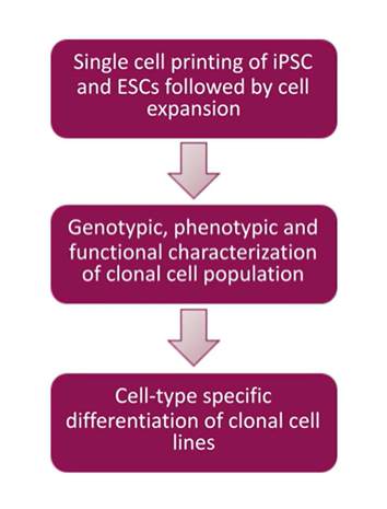
A simple workflow to start with any cell population, including your parental cell, and used to clone something out or an engineered cell to make cells any desired property.


Challenges in single-cell isolation and clonal outgrowth

Even with cutting-edge single-cell isolation technologies and monoclonality workflows, it’s still possible to run into some challenges.

According to Gargi Roy, the main challenge arose for her team when they wanted to print cells in multiple plates and form multiple colonies with the same characteristics. As the number of single-cell isolation repeats increases, one often observes a steady drop in clonal outgrowth efficiency. The decline is even sharper when you want to implement animal component-free (ACF) conditions, which you need to consider if animal-free research is critical to your institution.

The discrepancies in clonal outgrowth efficiency are more prominent in ESCs than in iPSCs. The efficiency drops down to single digits with engineered ESCs. The main reason for the decline is the drop in cell viability during plating, Roy states. She also emphasizes improving the experimental design by enriching the medium, especially in ACF conditions.

Colony Outgrowth Efficiencies from Single Cells between IPSCs and ESCs

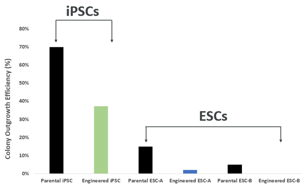
The differences in colony outgrowth efficiencies from single cells between IPSCs and ESCs illustrate significant discrepancies in ESCs.


Even when there is sufficient clonal outgrowth, you must take great care to prevent apoptosis. A timely harvest of 8-10 days is essential. It would help to feed your colony daily to prevent starvation.

Aggregation of the cells is another issue that hinders the monoclonality evaluation. You must use the correct dissociation reagent to detach cells from the well surface and from one another, which is a critical step for downstream analysis.


Assurance difficulties during monoclonal cell line development

Verification of monoclonality is crucial for cell line development. You need to provide detailed proof that your cell population is derived from a single cell for regulatory filing. Roy’s team found ESCs particularly hard to monitor because of their sticky surface and irregular shapes. If two ESCs in close proximity were dispensed, they would likely be detected as a single cell. Also, if you are facing a tight deadline, you could apply limiting dilution for statistics to calculate the chance of monoclonality.

CloneSelect Single-Cell Printer boasts advanced imaging techniques to prove monoclonality at the point of single-cell isolation. Not only can the printer easily distinguish between two very close cells during cell deposition, but it also allows you to trace back your colony from the day of harvest to day 0 to assure monoclonality as seen in the figure below.

single cell images with the CloneSelect Single-Cell Printer

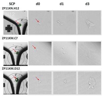
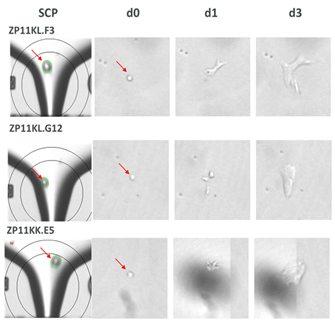
Confirmation of monoclonality and capturing single cell images with the CloneSelect Single-Cell Printer


Clonal characterization

The final component of cell line development is characterization. Once monoclonality has been assessed, you ensure this monoclonality is reflected in the genotype and pluripotency. Do the cells in your colony all have consistent levels of the desired functionality? More importantly, are they genomically stable, meaning they can conserve and transfer genetic material from one generation to the next? Lastly, are they pluripotent (i.e., can they differentiate into three main germ layers - ectoderm, endoderm, and mesoderm)? The answers are only obtainable by high-throughput clone screening.

This is where the CloneSelect Imager can add value to your research through additional confidence in monoclonality assurance. The CloneSelect Imager contains label-free brightfield technology with customizable fluorescence imaging, accompanied by advanced data analysis tools, including confluence measurements, growth curves, monoclonality report generation, and plate thumbnails to quickly visualize colony outgrowth in every well.

With the high resolution and variety of imaging modes, you can even detect the cell markers specific to the cells in your clone. This allows you to confirm if your monoclonal cell lines have the desired functionality.

The illustration below demonstrates the differentiation of monoclonal ESCs into three distinct germ layers, bearing the respective biomarkers in each layer.

High-quality/stable cells using our single cell printer

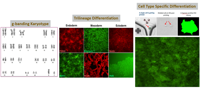
High-quality/stable cells using our single cell printer, making it easy to differentiate into three germ layers: endoderm, mesoderm, and ectoderm.


The CloneSelect Single-Cell Printer is an effective tool for single-cell isolation of iPSCs and ESCs. A combination of the CloneSelect Imager and the CloneSelect printer offers image-based proof of monoclonality assurance.

– Gargi Roy, Ph.D. Cell Culture & Fermentation Sciences of Bio-Pharmaceutical Development, AstraZeneca


The CloneSelect Imager’s scope of functional assessment continues even after you begin the cell differentiation stage. Through fluorescence imaging, you can monitor cells as they differentiate to form different tissue layers.


How can I learn more about monoclonal cell lines?

From the initial plating phase to outgrowth and cell differentiation, monoclonal cell line development is simpler using our CloneSelect workflow for Monoclonal assurance.

You can further explore the AstraZeneca Stem Cell Team's research for more information on the CloneSelect Single-Cell Printer and CloneSelect Imager monoclonality assurance workflow. In the recent Molecular Devices’ webinar, Gargi Roy illustrates the challenges and solutions in monoclonality assurance with details from her research that demonstrate how to engineer monoclonal cell lines and evaluate the cells in your colony.


CloneSelect Imager Monoclonality Assurance Workflow

Register today


Join the on-demand webinar, Monoclonality Assurance Using CloneSelect Single-Cell Printer and CloneSelect Imager in Human Stem Cell (hSC) Engineering Workflow, with Dr. Roy.


  1. Roy, Gargi, et al. "Sequential screening by ClonePix FL and intracellular staining facilitate isolation of high producer cell lines for monoclonal antibody manufacturing." Journal of immunological methods 451 (2017): 100-110.
  2. Frank, Richard T et al. “Concise review: stem cells as an emerging platform for antibody therapy of cancer.” Stem cells (Dayton, Ohio) vol. 28,11 (2010): 2084-7. doi:10.1002/stem.513.
  • Share on LinkedIn
  • Share on Facebook
  • Share on Twitter
  • Share on Youtube

Recent posts

View All Posts
Spacer

How 3D Cell Models Will Shape the Future of Drug Discovery

Target discovery and drug development rely heavily on 2D cell and animal…

A roundtable discussion
Spacer

A case study for assay-ready patient-derived organoids (PDOs) and high-throughput 3D imaging to advance drug discovery

Introduction – the problem.  The average cost of bringing a new drug to…

Read case study
Spacer

3D organoids and automation of complex cell assays [Podcast]

As we enter the era of sophisticated drug discovery with gene therapy and…

Discover 3D organoids
Spacer

Celebrating the art of science: A D+I interview with Daniele Tortorella

At Molecular Devices, we cultivate a culture where associates around the…

Celebrate D+I
Spacer

Advanced technology for automated 3D biology workflows #SLASEurope2022

SLAS Europe 2022, hosted numerous sessions packed with the latest…

View SLAS EU activities
Compare /

Microplate Reader

  • SpectraMax i3x
  • SpectraMax iD3 / iD5
  • SpectraMax M 시리즈
  • SoftMax Pro 소프트웨어

세포 이미징

  • ImageXpress Confocal HT.ai
  • ImageXpress Pico
  • IN Carta 이미지 분석
    소프트웨어

클론 스크리닝

  • ClonePix 2 포유동물 콜로니 피커
  • QPix 미생물 콜로니 피커

FLIPR System

AXON PATCH-CLAMP

실험실 자동화

GxP 컴플라이언스

  • 뉴스
  • 이벤트
2023년 4월 2일

Focus on Microscopy (FOM)

2023년 5월 9일

Imaging User Meeting 2023

2023년 6월 26일

MPS World Summit

행사 더 보기

2023년 2월 22일

Silver Sponsor Molecular Devices at Society for Laboratory Automation and Screening 2023 International Conference and Exhibition

2023년 2월 13일

Molecular Devices inks deal with HUB Organoids to advance automated intestinal organoid screening technology

2023년 2월 7일

Molecular Devices partners with SEED Biosciences to exclusively offer DispenCell Single-Cell Dispenser, expanding leadership in cell line development

뉴스 더 보기
뉴스레터 로더
  • 2023년 3월  
  • 2023년 2월  
  • 2023년 1월  

글로벌 본사

  • 주소 아이콘

    Molecular Devices, LLC.
    3860 N First Street
    San Jose, CA 95134

  • 연락처

    Phone: +1 800-635-5577

    Phone: +86-400-821-3787

    Phone: +44-118-944-8000

    Phone: +44-118-944-8000

    전화: +82-2-3471-9531

    Teléfono: +44-118-944-8000

    Telefono: +44-118-944-8000

  • 사무실 영업 시간

    Mon-Fri 8:00 am - 5:00 pm (PST)

    周一至周五,早上 9 点至下午 5 点 30 分

    Montag bis Freitag 8 h à 17 h (GMT)

    lundi à vendredi 8 h à 17 h (GMT)

    월~금, 오전 9시~오후 5시(한국표준시)

    de lunes a viernes 8 h à 17 h (GMT)

    lunedi al venerdì 8 h à 17 h (GMT)

  • 제품 영업

    Sales +1 877-589-2214

    售前咨询 : 400-821-3787 转 1

    Verkauf 00800 665 32860

    Ventes 00800 665 32860

    판매 +82-2-3471-9531

    Ventas 00800 665 32860

    Saldi 00800 665 32860

  • 기술 지원

    Support +1 800 635 5577

    售后咨询 : 400-821-3787 转 2

    Unterstützung +44-118-944-8000

    Support +44-118-944-8000

    지원 +82-2-3471-9531

    Apoyo +44-118-944-8000

    Supporto +44-118-944-8000

  • 자료 창고

    SpectraNet 고객 포털

바닥글 탐색

  • 이용약관
  • 개인정보 보호정책
  • 데이터를 판매하거나 공유하지 마십시오.
  • 온라인 이용약관
  • 상표
  • 지원
  • 연락처
  • 사이트맵
  • Danaher Life Sciences
  • 채용

©2023 Molecular Devices, LLC. 모든 권리 보유.

©2023 Molecular Devices, LLC. 美谷分子仪器(上海)有限公司保留所有权利。
All Rights Reserved 沪ICP备05056171号-1

barcode