메인 화면 바로가기
Molecular Devices, LLC
  •   美谷分子仪器(上海)有限公司
    咨询服务热线:400-821-3787
  •   0 Cart
  • Store

  •  채용
  •  언어  
    • English
    • Deutsch
    • 中文
    • 日本語
    • Français
    • 한국어
    • Italiano
    • Español
Molecular Devices, LLC
  • 제품
    • 제품

      새로운 DispenCell™ Single-Cell Dispenser 기술은 단일 세포주를 3배 더 빠르고 저렴한 비용으로 분리합니다.

      • 쉽고 직관적인 설정
      • 클론형성능과 추적 가능성의 즉각적인 증거
      • 고유한 기술로 세포 샘플을 부드럽게 처리
      • 벤치탑 크기  
        설계
      • 특허받은  
        일회용 팁
      DispenCell™ Single-Cell Dispenser 기술

      최적화된 3D 조직과 오가노이드 실험과정을 위한 BioAssemblyBot의 6축 로봇 팔을 갖춘 자동화된 High-Content Screening 솔루션

      BioAssemblyBot의 6축 로봇 팔

    • Microplate Reader
      SpectraMax Mini Multi-Mode Microplate Reader
      Multi-Mode Reader
      • SpectraMax i3x
      • SpectraMax iD3/iD5
      • SpectraMax M 시리즈
      • FlexStation 3
      • SpectraMax Mini
      SpectraMax ABS 마이크로플레이트
      흡광(Absorbance) 리더기
      • SpectraMax ABS/ABS Plus
      • SpectraMax VersaMax
      • SpectraMax QuickDrop
      • CMax Plus
      Fluorescence Reader
      Fluorescence Reader
      • SpectraMax Gemini
      SpectraMax Luminescence
      발광(Luminescence) 리더기
      • SpectraMax L


      MultiWash+ Washer
      Stacker 및 Washer
      • StakMax Stacker
      • AquaMax Washer
      • MultiWash+ Washer
      • MultiWash–C 微孔板洗板机
      SoftMax Pro 데이터 획득
      분석 소프트웨어
      • SoftMax Pro 소프트웨어
      • SoftMax Pro GxP Software
      GxP 솔루션
      GxP Compliance Solution
      • SoftMax Pro GxP Software
      • 소프트웨어 설치와 Validation 서비스
      • IQ/OQ/PM 서비스
      • SpectraTest Validation Plate
      실험실 자동화와 맞춤화
      실험실 자동화와 맞춤화
      • 플레이트 기반 High-Throughput Assay를 위한 실험실 자동화
    • 세포 Imaging 시스템
      ImageXpress Pico Automated Cell Imaging System
      Automated Cell Imaging Systems
      • ImageXpress Pico
      • ImageXpress Nano
      High-Content Imaging
      High-Content Imaging
      • ImageXpress Confocal HT.ai
      • ImageXpress Micro Confocal
      • ImageXpress Micro 4
      StratoMineR 분석
      Acquisition & Analysis Software
      • IN Carta
      • StratoMineR
      • MetaXpress
      • CellReporterXpress
      • MetaMorph
      실험실 자동화와 맞춤화
      실험실 자동화와 맞춤화
      • High-Throughput, High-Content Screening(HCS)을 위한 실험실 자동화
      • BioAssemblyBot 400 Bioprinter 자동화 HCS 솔루션
    • 클론 스크리닝
      Clone Pix 시리즈
      포유류 콜로니 피킹
      • ClonePix 2
      QPix 미생물 콜로니 피커
      미생물 콜로니 피킹
      • QPix 420
      • QPix 450/460
      • QPix HT
      CloneSelect Imager FL
      단일 세포 Imaging
      • CloneSelect Imager
      • CloneSelect Imager FL
      DispenCell Single-Cell Dispenser
      단일 세포 분리
      • DispenCell Single-Cell Dispenser
      실험실 자동화
      실험실 자동화와 맞춤화
      • High-Throughput 클론 스크리닝을 위한 실험실 자동화
      CloneMedia와 XP Media
      배지 및 시약
      Clone Screening Assay kit
      Clone Screening Assay kit
    • FLIPR Penta
      FLIPR Penta
      FLIPR Penta
      • FLIPR Penta High-Throughput 세포 스크리닝 시스템
      Screenworks
      분석 소프트웨어
      • ScreenWorks 소프트웨어
      • Peak Pro 2 소프트웨어 모듈
      Flipr Assay 키트
      FLIPR Assay 키트
      • 칼슘 Assay 키트
      • 칼륨 분석 키트
      • 막전위 분석키트
      • EarlyTox 심장 독성 키트
    • Axon Patch-Clamp
      투명
      Amplifier
      • Axopatch 200B 콘덴서
      • MultiClamp 700B
      • Axoclamp 900A
      투명
      Digitizer
      • Axon Digidata 1550B Low
      투명
      분석 소프트웨어
      • pCLAMP 11 소프트웨어 제품군
    • 기타
      Threshold Immunoassay System
      Threshold Immunoassay System
      Geneppix 미세배열 스캐너
      Genepix 미세배열 스캐너
      Imagexpress Micro Xls
      Imagexpress Micro xls
      인증 리퍼브
      인증 리퍼브
      IDBS 솔루션
      IDBS R&D 클라우드 솔루션
    • 분석 시약(assay kit)
      심장 독성
      • EarlyTox 심장 독성 키트
      Cell viability
      • EarlyTox Cell Integrity Kit
      • EarlyTox Cell Viability Assay Kit
      DNA 정량 분석
      • Spectramax Quant dsDNA Assay 키트
      Elisa, western blot
      • CatchPoint SimpleStep ELISA 키트
      • ScanLater Western Blot Assay 키트
      gpcr
      • FLIPR 칼슘 Assay 키트
      • Fura-2 QBT 칼슘 키트
      • CatchPoint cAMP 형광 Assay 키트
      • CatchPoint cGMP 형광 Assay 키트
      이온 통로
      • FLIPR 칼륨 Assay 키트
      • FLIPR 막전위 Assay 키트
      IGG 정량분석
      • ValitaTiter
      • CloneDetect
      Reporter gene
      • Spectramax Glo Steady-Luc Reporter Assay Kit
      • Spectramax DuoLuc Reporter Assay Kit
      Transporter
      • QBT Fatty Acid Uptake Assay Kit
      • 신경전달물질 수송체 흡수 Assay 키트
      기타
      • Contamination Detection
      • Enzyme - IMAP Assay
    • 액세서리 및 소모품
      Microplate Reader
      • 384 Well SBS
      • 384 Well High Sample Recovery Plate
      • Deep-well Plate
      • Low Profile Microplate
      • SpectraDrop Micro-Volume Microplate
      • SpectraMax Injection cartridge with SmartInject Technology
      • SpectraMax MiniMax 300
        Imaging Cytometer
      • Western Blot Cartridge
      • 96孔微孔板
      클론 스크리닝
      • Adjustable Petri Dish and Microplate Holder
      • Bioassay QTrays
      • Calibead
      • 캡 매트 및 뚜껑
      • 크로마 필터
      • 세정 및 살균 솔루션
      • CloneSelect Single-Cell Printer Cartridge
      • QPix 핀 및 헤드
      • QReps 레플리케이터
      Axon Patch-Clamp
      • Soft Panel Amplifier Control
      Spectra Img
  • 연구 분야
    • 응용 분야

      Molecular Devices, Cellesce 인수로 환자 유래 오가노이드 특허 기술 추가

      2022년 12월 6일

      • Cellesce의 동종 업계 최초 기술로 대규모 약물 Screening을 위한 일관적인 환자 유래 오가노이드를 생성합니다.
      • 인수를 통해 Molecular Devices의 3D 생물학 솔루션 혁신 기업으로서의 입지를 강화합니다.
      • 결합된 전문 지식으로 신약 개발을 위해 생리학적으로 연관된 세포 모델의 채택을 업계에서 가속화할 것입니다.


      OIC 방문하기

      언론 보도 읽기

      Cellsce
      응용 분야를 위한 Spectra
    • 코로나바이러스 (COVID-19)
      코로나19
      코로나19 연구
      관련 솔루션
      코로나19
      코로나19 관련
      새로운 뉴스
      코로나19
      백신 개발 워크플로
      감염병 연구 응용 분야
      백신 연구
      연구 응용 분야
    • 연구분야 (Stem Cell, Cancer)
      투명
      3D Cell Model
      투명
      암 연구 솔루션
      투명
      Cell Line Development
      투명
      신약 개발
      투명
      식품 및 음료
      투명
      유전자 편집(CRISPR/Cas9)
      투명
      오가노이드 연구
      투명
      줄기세포 연구
      투명
      독성학
    • Microplate Reader
      투명
      세포 건강 상태 (Cell Health)
      투명
      Cellular Signaling
      투명
      ELISA
      투명
      미생물학과 오염물질
      투명
      핵산(DNA/RNA) 측정과 분석
      투명
      단백질 측정, 정량분석, 분석
      투명
      관련 분석법: 측정 모드
      • 흡광(Absorbance)
      • 형광(Fluorescence)
      • 형광 편광
      • 발광(Luminescence)
      • TRF, TR-FRET 및 HTRF
      • Western Blot
    • 세포 Imaging 시스템
      투명
      Cell Counting
      투명
      세포 이미징 및 분석
      투명
      세포 Migration Assay
      투명
      세포 염색
      투명
      Live Cell Imaging
      투명
      Neurite Outgrowth
      투명
      장기 칩
      투명
      오가노이드
      투명
      Spheroids
    • 클론 스크리닝
      투명
      Cell line development 실험과정
      투명
      단클론항체(mAb)
      • 하이브리도마
      • 파지 디스플레이
      • 단일클론항체 생산
      투명
      Monoclonality
      투명
      합성 생물학
    • FLIPR Penta
      투명
      GPCR(G protein-coupled receptor)
      투명
      이온 채널
      투명
      Cardiotoxicity(심장독성)
    • Axon Patch-Clamp
      투명
      전기생리학(Patch Clamp)
  • 자료
    • 자료
    • 관련 자료 검색
      메뉴 자료 아이콘/응용 분야 노트
      Application Note
      메뉴 자료 아이콘/레퍼런스
      레퍼런스
      Ebook 아이콘
      eBook
      메뉴 자료 아이콘/Scientific Poster
      Scientific Poster
      메뉴 자료 아이콘/튜토리얼 및 영상
      동영상 및 웨비나

      검색

    • 블로그 – 실험실 노트
      스페이서
      Assay에 대한 고객 사례…
      스페이서
      3D organoids and…
      How 3D Cell Models Will Shape the Future of Drug Discovery
      2023년 3월 7일 Target discovery and drug development rely heavily on 2D cell and animal models to decipher efficacy and toxic effect of drug candidates. Yet, 90% of candidates fail to…
      더 알아보기  
    • 고객 사례 소개

      다른 연구자들이 제품과 솔루션을
      어떻게 사용했는지 확인해보세요.

    • 혁신 기술
      투명
      AI, 머신러닝 및 딥러닝
      투명
      AgileOptix 스피닝 디스크 기술
      투명
      자동 초점
      투명
      디지털 공초점 옵션
      투명
      고함량 Screening
      투명
      HumSilencer
      투명
      레이저 조명
      투명
      QuickID 표적 이미지 획득
    • 관련 분석법
      투명
      흡광(Absorbance)
      투명
      전기생리학
      투명
      형광(Fluorescence)
      투명
      형광 편광(FP)
      투명
      발광(Luminescence)
      투명
      TRF, TR-FRET 및 HTRF
      투명
      Water Immersion Objective
      투명
      Western Blot
    • 동영상 갤러리
      투명
      Microplate Reader
      투명
      세포 Imaging 시스템
      투명
      Flipr 시스템
      투명
      클론 스크리닝
      투명
      Axon Patch-Clamp
      투명
      주문형 웨비나
  • 서비스 및 지원
    • 서비스 및 지원
    • 개요
      Spectra 로고
      SpectraNet 고객 관리 포털
      GxP 컴플라이언스
      GxP 컴플라이언스 솔루션
      실험실 자동화와 맞춤화
      실험실 자동화와 맞춤화
      전문 서비스
      전문 서비스

      기술 지원

      미국 본사  
      +1 800-635-5577  
      월~금, 오전 7시~오후 5시 PST

      유럽  
      +44-118-944-8000  
      월~금, 오전 8시~오후 5시 GMT

      내가 있는 지역의 담당자 확인하기

      SpectraNet 고객 포털  

    • 고객 포털 - Spectranet
      Spectranet

      INTRODUCING OUR NEW CUSTOMERCARE PORTAL

      SpectraNet is an intuitive, simple-to-use, self-service customer portal providing a new level of experience available 24/7.

      Create your account today to get full access to integrated content and world-class customer service.

      REGISTER LOGIN
      지원 티켓 신청
      지원 티켓 신청하기
      자료 창고
      지식 기반
      제품 등록
      제품 등록하기
      기술 자료
      기술 자료
    • GxP 컴플라이언스 솔루션
      GxP Softmax Pro GxP 소프트트웨어
      SOFTMAX PRO GXP 소프트웨어
      GxP 소프트웨어 설치
      소프트웨어 설치와 Validation 서비스
      GxP Spectratestt Validation Plate 재인증
      SPECTRATEST Validation Plate
      IQ OQ 서비스
      IQ/OQ/PM 서비스
    • 실험실 자동화
      실험실 자동화와 맞춤화
      실험실 자동화와 맞춤화
      High Content Screening HCS
      High-Throughput, High-Content Screening
      • BioAssemblyBot 400 Bioprinter 자동화 HCS 솔루션
      플레이트 기반 High-Throughput Assay
      플레이트 기반 High-Throughput Assay
      High-Throughput 클론 스크리닝
      High-Throughput 클론 스크리닝
    • 전문 서비스
      전문 서비스
      전문 서비스
      서비스 플랜
      서비스 플랜
  • 회사
    • 회사

      Molecular Devices, 오스트리아의 글로벌 R&D 허브 확장

      2022년 10월 12일        
      더 큰 부지는 Cell Line Development, 오가노이드 개발, 신약 개발을 개선하기 위한 Screening 솔루션을 발전시키기 위한 협업 공간인 잘츠부르크의 오가노이드 혁신 센터의 미래 기지가 될 것입니다.


      OIC 방문하기

      언론 보도 읽기

      Austrian Research & Development Center의 리본 커팅 기념식
      응용 분야를 위한 Spectra
    • 회사 소개

      Molecular Devices는 실리콘 밸리를 기반으로 제약 및 연구의 스크리닝과 효과적인 분석이 가능한 솔루션을 40년에 걸쳐 개발하고 공급해 왔습니다.

    • 리더십

      당사의 다양한 경험, 비즈니스 인사이트 및 공동의 목표의식은 직원들이 잠재력을 최대한 발휘하도록 독려하기 위한 당사의 일상적인 결정에 추진력을 부여합니다.

      리더십

    • 채용

      당사의 팀 중심 기업 문화는 생각과 관점의 다양성과 강력한 신뢰 관계를 보장합니다.

    • 뉴스룸
      투명
      뉴스
      투명
      보도 내용
      Silver Sponsor Molecular Devices at Society for Laboratory Automation and Screening 2023 International Conference and Exhibition
      Feb 22, 2023 Showcasing new industry collaborations, automated technology, and workflow innovations that span 3D biology, cell line development, and drug…
      Read more  
    • 이벤트
      Focus on Microscopy (FOM)
      Conference | Europe | Porto, Portugal, Europe– Apr 02 – Apr 5, 2023 FOM2023 continues a long-standing (since 1988), yearly conference series on the latest innovations and developments in (optical) microscopy and their…
      Read more  
      Imaging User Meeting 2023
      Conference | Europe | Copenhagen, Denmark– May 09 – May 10, 2023 Ideal for both current ImageXpress system users and those wanting to learn more about high-content, high-throughput, automated or 3D imaging, our…
      Read more  
  • 연락처
  • 검색

    검색

  • 견적
    요청하기

모바일 탐색기

  • 제품
    • 모든 제품
    • Microplate Reader
      • Multi-Mode Reader
      • 흡광(Absorbance) 리더기
      • Fluorescence Reader
      • 발광(Luminescence) 리더기
      • Microplate Stacker
      • Microplate Washer
      • SoftMax Pro 소프트웨어
      • SoftMax Pro GxP Software
      • GxP Compliance Solution
      • 실험실 자동화와 맞춤화
    • 세포 Imaging 시스템
      • Automated Cell Imaging Systems
      • High-Content Imaging
      • Acquisition & Analysis Software
      • 실험실 자동화와 맞춤화
    • 클론 스크리닝
      • 포유류 콜로니 피킹
      • 미생물 콜로니 피킹
      • 단일 세포 Imaging
      • 단일 세포 분리
      • 실험실 자동화와 맞춤화
      • 배지 및 시약
      • Clone Screening Assay kit
    • FLIPR Penta
      • FLIPR Penta
      • 분석 소프트웨어
      • Flipr Assay 키트
    • Axon Patch-Clamp
      • Amplifier
      • Digitizer
      • pCLAMP 소프트웨어
    • 기타
      • Threshold Immunoassay System
      • GenePix 마이크로어레이 시스템
      • ImageXpress Micro XLS
      • 인증 리퍼브
      • IDBS R&D 클라우드 솔루션
    • 분석 시약(assay kit)
      • 심장 독성
      • Cell Viability
      • DNA 정량분석
      • ELISA, Western Blot
      • GPCR
      • 이온채널
      • IgG 정량분석
      • Reporter Gene
      • 수송체
      • 기타
    • 액세서리 및 소모품
      • Microplate Reader
      • 클론 스크리닝
      • Axon Patch-Clamp
  • 연구 분야
    • 모든 응용 분야
    • 코로나바이러스 (COVID-19)
      • 코로나19 연구
        관련 솔루션
      • 코로나19 관련
        새로운 뉴스
      • 백신 개발 워크플로
      • 백신 연구
        연구 응용 분야
    • 연구분야 (Stem Cell, Cancer)
      • 3D Cell Model
      • 암 연구 솔루션
      • Cell Line Development
      • 신약 개발
      • 식품 및 음료
      • 유전자 편집(CRISPR/Cas9)
      • 오가노이드 혁신 센터
      • 줄기세포 연구
      • 독성학
    • Microplate Reader
      • 세포 건강 상태 (Cell Health)
      • Cellular Signaling
      • ELISA
      • 미생물학과 오염물질
      • 핵산(DNA/RNA) 측정과 분석
      • 단백질 측정, 정량분석, 분석
      • 관련 분석법: 측정 모드
        • 흡광(Absorbance)
        • 형광(Fluorescence)
        • 형광 편광
        • 발광(Luminescence)
        • TRF, TR-FRET 및 HTRF
        • Western Blot
    • 세포 Imaging 시스템
      • Cell Counting
      • 세포 이미징 및 분석
      • 세포 Migration Assay
      • 세포 염색
      • Live Cell Imaging
      • Neurite Outgrowth
      • 장기 칩
      • 오가노이드
      • Spheroids
    • 클론 스크리닝
      • 단클론항체(mAb)
      • Monoclonality
      • 합성 생물학
    • FLIPR Penta
      • GPCR(G protein-coupled receptor)
      • 이온 채널
      • Cardiotoxicity(심장독성)
    • Axon Patch-Clamp
      • 전기생리학(Patch Clamp)
  • 자료
    • 관련 자료 검색
      • Application Note
      • 레퍼런스
      • eBook
      • Scientific Poster
      • 동영상 및 웨비나
    • 블로그-실험실 노트
    • 고객 사례 소개
    • 혁신 기술
      • AI, 머신러닝 및 딥러닝
      • AgileOptix 스피닝 디스크 기술
      • 자동 초점
      • 디지털 공초점 옵션
      • 고함량 Screening
      • HumSilencer
      • 레이저 조명
      • QuickID 표적 이미지 획득
    • 관련 분석법
      • 흡광(Absorbance)
      • 전기생리학
      • 형광(Fluorescence)
      • 형광 편광(FP)
      • 발광(Luminescence)
      • TRF, TR-FRET 및 HTRF
      • Water Immersion Objective
      • Western Blot
    • 동영상 갤러리
      • Microplate Reader
      • 세포 Imaging 시스템
      • Flipr 시스템
      • 클론 스크리닝
      • Axon Patch-Clamp
      • 주문형 웨비나
  • 서비스 및 지원
    • 개요
      • SpectraNet 고객 관리 포털
      • GxP 컴플라이언스 솔루션
      • 실험실 자동화 솔루션
      • 전문 서비스
    • 고객 포털 - Spectranet
      • 등록
      • 로그인
      • 지원 티켓 신청하기
      • 지식 기반
      • 제품 등록하기
      • 기술 자료
    • GxP 컴플라이언스 솔루션
      • SOFTMAX PRO GXP 소프트웨어
      • 소프트웨어 설치와 Validation
      • SpectraTest Validation Plate
      • IQ/OQ/PM 서비스
    • 실험실 자동화
      • 실험실 자동화와 맞춤화
      • High-Throughput, High-Content Screening
      • 플레이트 기반 High-Throughput Assay
      • High-Throughput 클론 스크리닝
    • 전문 서비스
      • 전문 서비스
      • 서비스 플랜
  • 회사
    • 회사 소개
    • 리더십
    • 채용
    • 뉴스룸
      • 뉴스
      • 보도 내용
    • 이벤트
  • 연락처
  • 견적
    요청하기
  1. 홈
  2. Lab Notes
  3. 클론 스크리닝
  4. [팟캐스트] 기존 Cell Line Development의 도전과제와 Monoclonality 조절에 도움이 되는 신기술
Molecular Devices Lab Notes

[팟캐스트] 기존 Cell Line Development의 도전과제와 Monoclonality 조절에 도움이 되는 신기술

  • 2022년 9월 27일
  • Guest bloggers: Dr. Natasa Skoko & Dr. Hugh Graham

유전 공학 및 합성 생물학의 발전은 최근 수십 년 동안 수많은 혁신적 발견이 가능하도록 했습니다. The importance of cell line development needs an honorable mention. Without it, many life-saving vaccines for infectious diseases, antibody drugs, and recombinant protein products, including insulin would not be widely available The most challenging step of compliant cell line development is monoclonality assurance, primarily when carried out manually. Monoclonality of therapeutic cell lines must be accomplished and documented for regulatory reasons.

In our podcast, “Cell line development with Dr. Natasa Skoko, ICGEB & Dr. Hugh Graham, MacroGenics,” we discuss traditional workflows for cell line development and the emerging technologies to help verify monoclonality.

Here we’ve summarized key discussion points from expert speakers: Dr. Natasa Skoko of Group Leader of the Biotechnology Development Unit at the International Centre for Genetic Engineering and Biotechnology (ICGEB), and Dr. Hugh Graham, Director of Cell Culture Sciences at MacroGenics.

Cell Line Development

  • Significance of stable cell lines
  • Challenges of a traditional cell line development workflow

Monoclonality Assurance

  • Essentials of monoclonal cell line
  • Importance of verifying early monoclonality

Emerging Technologies and Regulatory Impact

  • Emerging monoclonal technologies to verify single cell lines
  • The role of regulation and its impact on the cell line development process
  • What does the future hold for monoclonal cell line workflows?
  • Automated cell line development workflow with monoclonality assurance

Significance of stable cell lines

In the simplest form, the term cell line defines a population of cells that can be maintained in a culture for a certain period and retain a distinct phenotype, function, and stability.

Dr. Skoko summarizes the historical significance of the stable cell line. “The use of the cell line made the revolution, not just in scientific research and helped in understanding many fundamental biological processes,” Its applications range from antibody and therapeutic protein production to drug screening.

Cell line development involves engineered mammalian cells that are robust, cost-effective, and easy to grow. One cell line that fits all the criteria is the Chinese Hamster Ovary (CHO) cell, first developed in 1987. Today, 70% of biotherapeutics on the market are manufactured from CHO cell lines. They are particularly efficient in the mass production of biotherapeutics, thanks to their adaptability to various growth media and growth conditions (e.g. suspension, attached growth, fed batch, perfusion) as well as their ability to fold and glycosylate proteins to mimic post-translational modifications in human proteins.

Challenges of a traditional cell line development workflow

The production of high levels of biotherapeutics relies on the generation of stable cell lines.

The first step is transfection of a suitable host cell line with the gene of interest, leading to random integration of DNA into the host genome. Once the engineered cell line starts growing, usually under selection pressure, single-cell isolation is needed to generate clones for screening.

In traditional workflows, cells are plated onto a 96-well plate through the limiting dilution. This method involves the dilution of the cell suspension such that when you plate a certain amount into a well, the likelihood of it having only one cell is very high. Some wells can even be empty and a few may have multiple cells. Therefore, you have a high probability that a cell line from any one of these wells is developed from a single cell.

This is great, but how long does it take? Assuming cells double in numbers every 24 hours, it takes ten doublings to reach a thousand cells, which would take 10 days. To obtain enough cells to work with, would take 2-3 weeks. Moreover, regulations for monoclonality require that you repeat limiting dilution to have further assurance of clonal origin for your cell line. So, you would need to spend months to obtain results from this low-throughput method. Dr. Graham adds: “The stability of the cell line is challenged. By the time you reach the desirable numbers and have high assurance of monoclonality, there is a chance they will have drifted in their properties, lost some copies of the gene of interest, or adapted in a way you didn’t expect.”

Automation is the way to facilitate high throughput monoclonal cell line growth and its documentation.

To sum up, limiting dilution can fail to achieve speed, compliance and cell line quality because it is hard to document monoclonality and retain the quality of your cells. Both Dr. Skoko and Dr. Graham agree that automation is the way to facilitate high throughput monoclonal cell line growth and its documentation.

Essentials of monoclonal cell line

Let’s now take a step back to define monoclonality. A monoclonal cell line originates from a single cell or from a single progenitor.

Why is it such a big deal? As cells start to grow and double, they are subject to genetic drifts, mutations, or loss of a plasmid. So, it is nearly impossible to ensure that the quality of the therapeutic proteins they express is uniform by simply investigating their most recent phenotype.

To compensate for that, you need to start documenting your cells on Day Zero to prove that they indeed came from a single cell.

Monoclonal Cell Line CHO-s cell growth

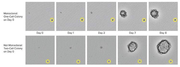
CHO-s cell growth was used to capture images from the 6-well plates at multiple time points. 0일 차에, 위 열에 하나의 세포가 존재한다는 것이 명확하게 관찰되었고, 아래 열에 두 개의 세포가 관찰되었습니다. The yellow circle shows the position of a bead that serves as a location reference to confirm that the same colony is imaged over time.

Typical monoclonality evidence that regulators look for is image-based, i.e., the image of a single cell must be recorded.

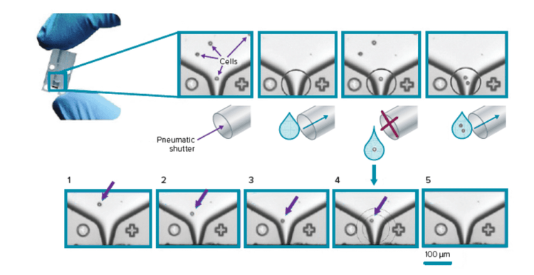
The most effective monoclonal cell line development equipment is a single cell printer, which performs single-cell deposition. Other methods include Fluorescence-Activated Cell Sorting (FACS) and workflows that will automate every step, from incubation to pipetting.

Importance of verifying early monoclonality

Monoclonality assurance at an early stage has several implications for life science applications. Dr. Graham highlights the significance of monoclonality for the drug development cycle: “There are cell lines and molecules going towards commercialization that were developed ten or more years ago. The cloning processes from ten years ago may not have the documentation required to assure a clonal origin. When one takes such a cell line to a regulator, you might be asked to show that it originally came from a clonal cell line. If you don’t have that proof, then proving it later in the project is a lot of work.”

“It’s all about minimizing risk and having a consistent production process and product quality. That’s why it’s essential to assure monoclonality at the early stage.”

Dr. Skoko adds: “It’s all about minimizing risk and having a consistent production process and product quality. That’s why it’s essential to assure monoclonality at the early stage. Starting with a cell bank with a high assurance of monoclonality results in less work later. Furthermore, you will avoid serious disruptions in manufacturing and reduce uncertainty when making changes to the manufacturing process.”

Emerging monoclonal technologies to verify single cell lines

Emerging techniques for monoclonal cell line imaging and cultivation are growing. Here are some examples:

  • Semi-solid cell suspension, whereby a single cell grows in a semi-solid suspension, can be imaged as it grows.
  • A cloning ring surrounding a single cell to isolate it and protect it from contamination
  • FACS: The sorting of cells based on fluorescent characteristics
  • Microfluidic drop-based single-cell printers with the ability to image newly-plated wells containing single cells

These methods combined improve the credibility of monoclonality evidence.

Of course, there is great potential to improve the current techniques and invent superior ones. First and foremost, visual evidence, i.e., single-cell imaging, must be used more often since it overcomes the cost and time-related challenges of limiting dilution and flow cytometry sorting.

CloneSelect 단일세포 프린터 시리즈

CloneSelect 단일 세포 프린터 시리즈는 완전 자동화 시스템으로, 단일 세포를 표준 Microplate에 정렬 및 배치하기 위해 독점 미세 유체공학 기반 기술과 실시간 이미지 분석을 활용하는 동시에 이미지 문서화로 Monoclonality를 보장합니다.

Further improvements are possible down the production line. For instance, improving analytical methods in therapeutic protein production can help us monitor the entire manufacturing process. This brings genetic variation under control and ensures uniformity of the manufactured protein.

Another concern in monoclonal cell line development is speed. As Dr. Graham points out, “Waiting for a clonal cell line to make your protein to go into a clinic is a barrier in terms of time. Today, people are moving towards the first batches of proteins from pools of early clones for the first Good Manufacturing Practice (GMP) clinical material. Then, one generates the clonal cell line for later-stage clinical trials and GMP production. Eliminating the need for proof of clonal origin in early clinical trials is an interesting approach to accelerate biotherapeutics development. However, this approach may need additional work to gain regulatory approval.”

The role of regulation and its impact on the cell line development process

As you can see, regulations undeniably impact monoclonal cell line development. One implication of stringent regulations is that the manufacturer must provide a comprehensive report on the monoclonal cell line before requesting approval. Insufficient proof or missing documents may result in regulatory delays or additional post-approval commitments. Because regulatory approval requires excellent scientific support, it is best to provide as much proof from the early stages as possible, especially for the clonal origin.

What does the future hold for monoclonal cell line workflows?

There are unresolved challenges in monoclonal cell line workflows, including financial concerns, obtaining high-throughput results, and assurance of monoclonal origin.

Compared with legacy methods, today’s monoclonal cell line developers have much better tools to ensure regulatory compliance and cell line quality.

One exciting prospect is improved data management, which is necessary to organize large datasets generated from cell line development studies. Combining data management with machine learning can give rise to a platform that predicts the quality of a biotherapeutic based solely on clonal origin.

Automated technology can help us have accurate clones, eliminate errors associated with the traditional limiting dilution, and overcome working with a heterogeneous population of different secretors.

The role of automation will also become more pivotal in speed and consistency. More specifically, automation can accelerate clone development and colony screening. Dr. Skoko clarifies the effects of automation: “We are talking about screening 10,000 clones in a couple of weeks. A traditional workflow would take anywhere from 30 weeks to two years. Automated technology can help us have accurate clones, eliminate errors associated with the traditional limiting dilution, and overcome working with a heterogeneous population of different secretors. You can evaluate the stability of your clones from the very beginning.”

Automated cell line development workflow with monoclonality assurance

With the integration of automation technologies, you can screen hundreds of thousands of clones to find that golden clone that can generate the highest yield for the protein of your interest. More importantly, it helps you fully document every step of the development process from day zero. Ultimately, you get to save money and time by avoiding future conflicts caused by a lack of monoclonality assurance or inconsistent product quality.

Cell Line Development Workflow for Monoclonality Assurance

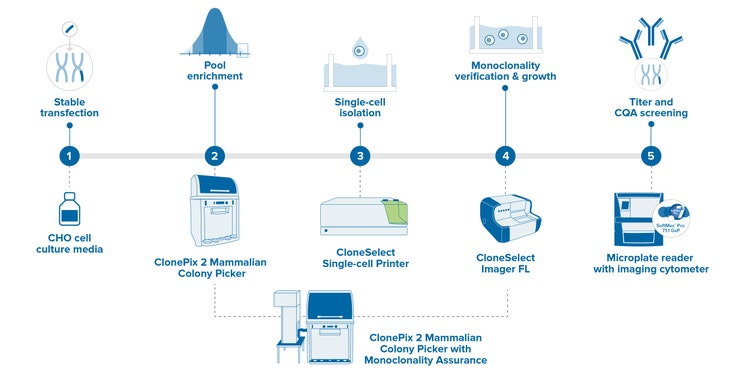
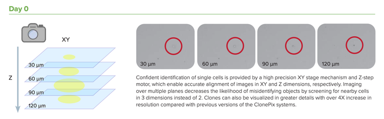
Accelerate your cell line development timelines by integrating multiple steps into a single step. The enhanced ClonePix 2 Mammalian Colony Picker with monoclonality assurance can automatically screen and pick clones that are both high producing and monoclonal—all in one system. Screen more clones in less time with monoclonal verification on day zero, then screen and pick for highest produces in less than two weeks. Rapid Z-stack acquisition feature allows detection of single cells throughout the medium volume, not just a single focal plane.

Cell Line Development timelines steps

Molecular Devices has a robust portfolio to automate your cell line development workflow and technologies to assure monoclonality, including clone picking, single-cell isolation and imaging, and microplate readers. Our proven GxP solutions help assure data integrity and compliance for GMP/GLP labs.

Learn more about cell line development and monoclonality applications or speak to an automation specialist if you’re interested in exploring an automated solution for your lab.

  • Share on LinkedIn
  • Share on Facebook
  • Share on Twitter
  • Share on Youtube

Recent posts

View All Posts
스페이서

How 3D Cell Models Will Shape the Future of Drug Discovery

Target discovery and drug development rely heavily on 2D cell and animal…

A roundtable discussion
스페이서

Assay에 사용할 준비가 된 환자 유래 오가노이드(PDO) 및 신약 개발 진전을 위한 High-Throughput 3D Imaging에 관한 고객 사례

Introduction – the problem.  The average cost of bringing a new drug to…

Read case study
스페이서

복잡한 세포 Assay의 3D 오가노이드 및 자동화[팟캐스트]

As we enter the era of sophisticated drug discovery with gene therapy and…

Discover 3D organoids
스페이서

과학의 아름다움을 기념하다. A D+I interview with Daniele Tortorella

At Molecular Devices, we cultivate a culture where associates around the…

Celebrate D+I
스페이서

자동화된 3D 생물학 실험과정을 위한 고급 기술 #SLASEurope2022

SLAS Europe 2022, hosted numerous sessions packed with the latest…

View SLAS EU activities
비교 /

Microplate Reader

  • SpectraMax i3x
  • SpectraMax iD3 / iD5
  • SpectraMax M 시리즈
  • SoftMax Pro 소프트웨어

세포 이미징

  • ImageXpress Confocal HT.ai
  • ImageXpress Pico
  • IN Carta 이미지 분석
    소프트웨어

클론 스크리닝

  • ClonePix 2 포유동물 콜로니 피커
  • QPix 미생물 콜로니 피커

FLIPR System

AXON PATCH-CLAMP

실험실 자동화

GxP 컴플라이언스

  • 뉴스
  • 이벤트
2023년 4월 2일

Focus on Microscopy (FOM)

2023년 5월 9일

Imaging User Meeting 2023

2023년 6월 26일

MPS World Summit

행사 더 보기

2023년 2월 22일

Silver Sponsor Molecular Devices at Society for Laboratory Automation and Screening 2023 International Conference and Exhibition

2023년 2월 13일

Molecular Devices inks deal with HUB Organoids to advance automated intestinal organoid screening technology

2023년 2월 7일

Molecular Devices partners with SEED Biosciences to exclusively offer DispenCell Single-Cell Dispenser, expanding leadership in cell line development

뉴스 더 보기
뉴스레터 로더
  • 2023년 3월  
  • 2023년 2월  
  • 2023년 1월  

글로벌 본사

  • 주소 아이콘

    Molecular Devices, LLC.
    3860 N First Street
    San Jose, CA 95134

  • 연락처

    Phone: +1 800-635-5577

    Phone: +86-400-821-3787

    Phone: +44-118-944-8000

    Phone: +44-118-944-8000

    전화: +82-2-3471-9531

    Teléfono: +44-118-944-8000

    Telefono: +44-118-944-8000

  • 사무실 영업 시간

    Mon-Fri 8:00 am - 5:00 pm (PST)

    周一至周五,早上 9 点至下午 5 点 30 分

    Montag bis Freitag 8 h à 17 h (GMT)

    lundi à vendredi 8 h à 17 h (GMT)

    월~금, 오전 9시~오후 5시(한국표준시)

    de lunes a viernes 8 h à 17 h (GMT)

    lunedi al venerdì 8 h à 17 h (GMT)

  • 제품 영업

    Sales +1 877-589-2214

    售前咨询 : 400-821-3787 转 1

    Verkauf 00800 665 32860

    Ventes 00800 665 32860

    판매 +82-2-3471-9531

    Ventas 00800 665 32860

    Saldi 00800 665 32860

  • 기술 지원

    Support +1 800 635 5577

    售后咨询 : 400-821-3787 转 2

    Unterstützung +44-118-944-8000

    Support +44-118-944-8000

    지원 +82-2-3471-9531

    Apoyo +44-118-944-8000

    Supporto +44-118-944-8000

  • 자료 창고

    SpectraNet 고객 포털

바닥글 탐색

  • 이용약관
  • 개인정보 보호정책
  • 데이터를 판매하거나 공유하지 마십시오.
  • 온라인 이용약관
  • 상표
  • 지원
  • 연락처
  • 사이트맵
  • Danaher Life Sciences
  • 채용

©2023 Molecular Devices, LLC. 모든 권리 보유.

©2023 Molecular Devices, LLC. 美谷分子仪器(上海)有限公司保留所有权利。
All Rights Reserved 沪ICP备05056171号-1

barcode