Molecular Devices, LLC
  •   美谷分子仪器(上海)有限公司
    咨询服务热线:400-821-3787
  •   0 Cart
  • Store

  •  채용
  •  언어  
    • English
    • Deutsch
    • 中文
    • 日本語
    • Français
    • 한국어
    • Italiano
    • Español
Molecular Devices, LLC
  • 제품
    • 제품

      새로운 DispenCell™ Single-Cell Dispenser 기술은 단일 세포주를 3배 더 빠르고 저렴한 비용으로 분리합니다.

      • 쉽고 직관적인 설정
      • 클론형성능과 추적 가능성의 즉각적인 증거
      • 고유한 기술로 세포 샘플을 부드럽게 처리
      • 벤치탑 크기  
        설계
      • 특허받은  
        일회용 팁
      DispenCell™ Single-Cell Dispenser 기술

      최적화된 3D 조직과 오가노이드 실험과정을 위한 BioAssemblyBot의 6축 로봇 팔을 갖춘 자동화된 High-Content Screening 솔루션

      BioAssemblyBot의 6축 로봇 팔

    • Microplate Reader
      SpectraMax Mini Multi-Mode Microplate Reader
      Multi-Mode Reader
      • SpectraMax i3x
      • SpectraMax iD3/iD5
      • SpectraMax M 시리즈
      • FlexStation 3
      • SpectraMax Mini
      SpectraMax ABS 마이크로플레이트
      흡광(Absorbance) 리더기
      • SpectraMax ABS/ABS Plus
      • SpectraMax VersaMax
      • SpectraMax QuickDrop
      • CMax Plus
      Fluorescence Reader
      Fluorescence Reader
      • SpectraMax Gemini
      SpectraMax Luminescence
      발광(Luminescence) 리더기
      • SpectraMax L


      MultiWash+ Washer
      Stacker 및 Washer
      • StakMax Stacker
      • AquaMax Washer
      • MultiWash+ Washer
      • MultiWash–C 微孔板洗板机
      SoftMax Pro 데이터 획득
      분석 소프트웨어
      • SoftMax Pro 소프트웨어
      • SoftMax Pro GxP Software
      GxP 솔루션
      GxP Compliance Solution
      • SoftMax Pro GxP Software
      • 소프트웨어 설치와 Validation 서비스
      • IQ/OQ/PM 서비스
      • SpectraTest Validation Plate
      실험실 자동화와 맞춤화
      실험실 자동화와 맞춤화
      • 플레이트 기반 High-Throughput Assay를 위한 실험실 자동화
    • 세포 Imaging 시스템
      ImageXpress Pico Automated Cell Imaging System
      Automated Cell Imaging Systems
      • ImageXpress Pico
      • ImageXpress Nano
      High-Content Imaging
      High-Content Imaging
      • ImageXpress Confocal HT.ai
      • ImageXpress Micro Confocal
      • ImageXpress Micro 4
      StratoMineR 분석
      Acquisition & Analysis Software
      • IN Carta
      • StratoMineR
      • MetaXpress
      • CellReporterXpress
      • MetaMorph
      실험실 자동화와 맞춤화
      실험실 자동화와 맞춤화
      • High-Throughput, High-Content Screening(HCS)을 위한 실험실 자동화
      • BioAssemblyBot 400 Bioprinter 자동화 HCS 솔루션
    • 클론 스크리닝
      Clone Pix 시리즈
      포유류 콜로니 피킹
      • ClonePix 2
      QPix 미생물 콜로니 피커
      미생물 콜로니 피킹
      • QPix 420
      • QPix 450/460
      • QPix HT
      CloneSelect Imager FL
      단일 세포 Imaging
      • CloneSelect Imager
      • CloneSelect Imager FL
      DispenCell Single-Cell Dispenser
      단일 세포 분리
      • DispenCell Single-Cell Dispenser
      실험실 자동화
      실험실 자동화와 맞춤화
      • High-Throughput 클론 스크리닝을 위한 실험실 자동화
      CloneMedia와 XP Media
      배지 및 시약
      Clone Screening Assay kit
      Clone Screening Assay kit
    • FLIPR Penta
      FLIPR Penta
      FLIPR Penta
      • FLIPR Penta High-Throughput 세포 스크리닝 시스템
      Screenworks
      분석 소프트웨어
      • ScreenWorks 소프트웨어
      • Peak Pro 2 소프트웨어 모듈
      Flipr Assay 키트
      FLIPR Assay 키트
      • 칼슘 Assay 키트
      • 칼륨 분석 키트
      • 막전위 분석키트
      • EarlyTox 심장 독성 키트
    • Axon Patch-Clamp
      투명
      Amplifier
      • Axopatch 200B 콘덴서
      • MultiClamp 700B
      • Axoclamp 900A
      투명
      Digitizer
      • Axon Digidata 1550B Low
      투명
      분석 소프트웨어
      • pCLAMP 11 소프트웨어 제품군
    • 기타
      Threshold Immunoassay System
      Threshold Immunoassay System
      Geneppix 미세배열 스캐너
      Genepix 미세배열 스캐너
      Imagexpress Micro Xls
      Imagexpress Micro xls
      인증 리퍼브
      인증 리퍼브
      IDBS 솔루션
      IDBS R&D 클라우드 솔루션
    • 분석 시약(assay kit)
      심장 독성
      • EarlyTox 심장 독성 키트
      Cell viability
      • EarlyTox Cell Integrity Kit
      • EarlyTox Cell Viability Assay Kit
      DNA 정량 분석
      • Spectramax Quant dsDNA Assay 키트
      Elisa, western blot
      • CatchPoint SimpleStep ELISA 키트
      • ScanLater Western Blot Assay 키트
      gpcr
      • FLIPR 칼슘 Assay 키트
      • Fura-2 QBT 칼슘 키트
      • CatchPoint cAMP 형광 Assay 키트
      • CatchPoint cGMP 형광 Assay 키트
      이온 통로
      • FLIPR 칼륨 Assay 키트
      • FLIPR 막전위 Assay 키트
      IGG 정량분석
      • ValitaTiter
      • CloneDetect
      Reporter gene
      • Spectramax Glo Steady-Luc Reporter Assay Kit
      • Spectramax DuoLuc Reporter Assay Kit
      Transporter
      • QBT Fatty Acid Uptake Assay Kit
      • 신경전달물질 수송체 흡수 Assay 키트
      기타
      • Contamination Detection
      • Enzyme - IMAP Assay
    • 액세서리 및 소모품
      Microplate Reader
      • 384 Well SBS
      • 384 Well High Sample Recovery Plate
      • Deep-well Plate
      • Low Profile Microplate
      • SpectraDrop Micro-Volume Microplate
      • SpectraMax Injection cartridge with SmartInject Technology
      • SpectraMax MiniMax 300
        Imaging Cytometer
      • Western Blot Cartridge
      • 96孔微孔板
      클론 스크리닝
      • Adjustable Petri Dish and Microplate Holder
      • Bioassay QTrays
      • Calibead
      • 캡 매트 및 뚜껑
      • 크로마 필터
      • 세정 및 살균 솔루션
      • CloneSelect Single-Cell Printer Cartridge
      • QPix 핀 및 헤드
      • QReps 레플리케이터
      Axon Patch-Clamp
      • Soft Panel Amplifier Control
      Spectra Img
  • 연구 분야
    • 응용 분야

      Molecular Devices, Cellesce 인수로 환자 유래 오가노이드 특허 기술 추가

      2022년 12월 6일

      • Cellesce의 동종 업계 최초 기술로 대규모 약물 Screening을 위한 일관적인 환자 유래 오가노이드를 생성합니다.
      • 인수를 통해 Molecular Devices의 3D 생물학 솔루션 혁신 기업으로서의 입지를 강화합니다.
      • 결합된 전문 지식으로 신약 개발을 위해 생리학적으로 연관된 세포 모델의 채택을 업계에서 가속화할 것입니다.


      OIC 방문하기

      언론 보도 읽기

      Cellsce
      응용 분야를 위한 Spectra
    • 코로나바이러스 (COVID-19)
      코로나19
      코로나19 연구
      관련 솔루션
      코로나19
      코로나19 관련
      새로운 뉴스
      코로나19
      백신 개발 워크플로
      감염병 연구 응용 분야
      백신 연구
      연구 응용 분야
    • 연구분야 (Stem Cell, Cancer)
      투명
      3D Cell Model
      투명
      암 연구 솔루션
      투명
      Cell Line Development
      투명
      신약 개발
      투명
      식품 및 음료
      투명
      유전자 편집(CRISPR/Cas9)
      투명
      오가노이드 연구
      투명
      줄기세포 연구
      투명
      독성학
    • Microplate Reader
      투명
      세포 건강 상태 (Cell Health)
      투명
      Cellular Signaling
      투명
      ELISA
      투명
      미생물학과 오염물질
      투명
      핵산(DNA/RNA) 측정과 분석
      투명
      단백질 측정, 정량분석, 분석
      투명
      관련 분석법: 측정 모드
      • 흡광(Absorbance)
      • 형광(Fluorescence)
      • 형광 편광
      • 발광(Luminescence)
      • TRF, TR-FRET 및 HTRF
      • Western Blot
    • 세포 Imaging 시스템
      투명
      Cell Counting
      투명
      세포 이미징 및 분석
      투명
      세포 Migration Assay
      투명
      세포 염색
      투명
      Live Cell Imaging
      투명
      Neurite Outgrowth
      투명
      장기 칩
      투명
      오가노이드
      투명
      Spheroids
    • 클론 스크리닝
      투명
      Cell line development 실험과정
      투명
      단클론항체(mAb)
      • 하이브리도마
      • 파지 디스플레이
      • 단일클론항체 생산
      투명
      Monoclonality
      투명
      합성 생물학
    • FLIPR Penta
      투명
      GPCR(G protein-coupled receptor)
      투명
      이온 채널
      투명
      Cardiotoxicity(심장독성)
    • Axon Patch-Clamp
      투명
      전기생리학(Patch Clamp)
  • 자료
    • 자료
    • 관련 자료 검색
      메뉴 자료 아이콘/응용 분야 노트
      Application Note
      메뉴 자료 아이콘/레퍼런스
      레퍼런스
      Ebook 아이콘
      eBook
      메뉴 자료 아이콘/Scientific Poster
      Scientific Poster
      메뉴 자료 아이콘/튜토리얼 및 영상
      동영상 및 웨비나

      검색

    • 블로그 – 실험실 노트
      스페이서
      Assay에 대한 고객 사례…
      스페이서
      3D organoids and…
      How 3D Cell Models Will Shape the Future of Drug Discovery
      2023년 3월 7일 Target discovery and drug development rely heavily on 2D cell and animal models to decipher efficacy and toxic effect of drug candidates. Yet, 90% of candidates fail to…
      더 알아보기  
    • 고객 사례 소개

      다른 연구자들이 제품과 솔루션을
      어떻게 사용했는지 확인해보세요.

    • 혁신 기술
      투명
      AI, 머신러닝 및 딥러닝
      투명
      AgileOptix 스피닝 디스크 기술
      투명
      자동 초점
      투명
      디지털 공초점 옵션
      투명
      고함량 Screening
      투명
      HumSilencer
      투명
      레이저 조명
      투명
      QuickID 표적 이미지 획득
    • 관련 분석법
      투명
      흡광(Absorbance)
      투명
      전기생리학
      투명
      형광(Fluorescence)
      투명
      형광 편광(FP)
      투명
      발광(Luminescence)
      투명
      TRF, TR-FRET 및 HTRF
      투명
      Water Immersion Objective
      투명
      Western Blot
    • 동영상 갤러리
      투명
      Microplate Reader
      투명
      세포 Imaging 시스템
      투명
      Flipr 시스템
      투명
      클론 스크리닝
      투명
      Axon Patch-Clamp
      투명
      주문형 웨비나
  • 서비스 및 지원
    • 서비스 및 지원
    • 개요
      Spectra 로고
      SpectraNet 고객 관리 포털
      GxP 컴플라이언스
      GxP 컴플라이언스 솔루션
      실험실 자동화와 맞춤화
      실험실 자동화와 맞춤화
      전문 서비스
      전문 서비스

      기술 지원

      미국 본사  
      +1 800-635-5577  
      월~금, 오전 7시~오후 5시 PST

      유럽  
      +44-118-944-8000  
      월~금, 오전 8시~오후 5시 GMT

      내가 있는 지역의 담당자 확인하기

      SpectraNet 고객 포털  

    • 고객 포털 - Spectranet
      Spectranet

      INTRODUCING OUR NEW CUSTOMERCARE PORTAL

      SpectraNet is an intuitive, simple-to-use, self-service customer portal providing a new level of experience available 24/7.

      Create your account today to get full access to integrated content and world-class customer service.

      REGISTER LOGIN
      지원 티켓 신청
      지원 티켓 신청하기
      자료 창고
      지식 기반
      제품 등록
      제품 등록하기
      기술 자료
      기술 자료
    • GxP 컴플라이언스 솔루션
      GxP Softmax Pro GxP 소프트트웨어
      SOFTMAX PRO GXP 소프트웨어
      GxP 소프트웨어 설치
      소프트웨어 설치와 Validation 서비스
      GxP Spectratestt Validation Plate 재인증
      SPECTRATEST Validation Plate
      IQ OQ 서비스
      IQ/OQ/PM 서비스
    • 실험실 자동화
      실험실 자동화와 맞춤화
      실험실 자동화와 맞춤화
      High Content Screening HCS
      High-Throughput, High-Content Screening
      • BioAssemblyBot 400 Bioprinter 자동화 HCS 솔루션
      플레이트 기반 High-Throughput Assay
      플레이트 기반 High-Throughput Assay
      High-Throughput 클론 스크리닝
      High-Throughput 클론 스크리닝
    • 전문 서비스
      전문 서비스
      전문 서비스
      서비스 플랜
      서비스 플랜
  • 회사
    • 회사

      Molecular Devices, 오스트리아의 글로벌 R&D 허브 확장

      2022년 10월 12일        
      더 큰 부지는 Cell Line Development, 오가노이드 개발, 신약 개발을 개선하기 위한 Screening 솔루션을 발전시키기 위한 협업 공간인 잘츠부르크의 오가노이드 혁신 센터의 미래 기지가 될 것입니다.


      OIC 방문하기

      언론 보도 읽기

      Austrian Research & Development Center의 리본 커팅 기념식
      응용 분야를 위한 Spectra
    • 회사 소개

      Molecular Devices는 실리콘 밸리를 기반으로 제약 및 연구의 스크리닝과 효과적인 분석이 가능한 솔루션을 30년에 걸쳐 개발하고 공급해 왔습니다.

    • 리더십
      Molecular Devices 리더십
    • 채용

      당사의 팀 중심 기업 문화는 생각과 관점의 다양성과 강력한 신뢰 관계를 보장합니다.

    • 뉴스룸
      투명
      뉴스
      투명
      보도 내용
      Silver Sponsor Molecular Devices at Society for Laboratory Automation and Screening 2023 International Conference and Exhibition
      Feb 22, 2023 Showcasing new industry collaborations, automated technology, and workflow innovations that span 3D biology, cell line development, and drug…
      Read more  
    • 이벤트
      Focus on Microscopy (FOM)
      Conference | Europe | Porto, Portugal, Europe– Apr 02 – Apr 5, 2023 FOM2023 continues a long-standing (since 1988), yearly conference series on the latest innovations and developments in (optical) microscopy and their…
      Read more  
      Imaging User Meeting 2023
      Conference | Europe | Copenhagen, Denmark– May 09 – May 10, 2023 Ideal for both current ImageXpress system users and those wanting to learn more about high-content, high-throughput, automated or 3D imaging, our…
      Read more  
  • 연락처
  • 검색

    검색

  • 견적
    요청하기

Mobile navigation

  • Products
    • All Products
    • Microplate readers
      • Multi-Mode Readers
      • Absorbance Readers
      • Fluorescence Readers
      • Luminescence Readers
      • Microplate Stacker
      • Microplate Washers
      • SoftMax Pro Software
      • SoftMax Pro GxP Software
      • GxP Compliance Solutions
      • Lab Automation & Customization
    • Cellular Imaging Systems
      • Automated Cell Imaging Systems
      • High-Content Imaging
      • Acquisition & Analysis Software
      • Lab Automation & Customization
    • Clone Screening
      • Mammalian Colony Picking
      • Microbial Colony Picking
      • Single-Cell Imaging
      • Single-cell Isolation
      • Lab Automation & Customization
      • Culture Media & Reagents
      • Clone Screening Assay kit
    • FLIPR Penta
      • FLIPR Penta
      • ANALYSIS SOFTWARE
      • FLIPR ASSAY KITS
    • Axon Patch-Clamp
      • Amplifiers
      • Digitizers
      • pCLAMP Software
    • Additional Products
      • Threshold Immunoassay System
      • GenePix Microarray Systems
      • ImageXpress Micro XLS
      • Certified Refurbished
      • R&D CLOUD SOLUTIONS BY IDBS
    • Assay Kits
      • Cardiotox
      • CELL VIABILITY
      • DNA QUANTITATION
      • ELISA, WESTERN BLOT
      • GPCR
      • ION CHANNEL
      • IgG quantitation
      • REPORTER GENE
      • Transporters
      • OTHER
    • Accessories and Consumables
      • MICROPLATE READERS
      • CLONE SCREENING
      • Axon Patch-Clamp
  • Applications
    • All Applications
    • SARS-CoV-2 (COVID-19)
      • COVID-19 Research Solution
      • COVID-19 Company Update
      • Vaccine Development Workflows
      • Vaccine Research Applications
    • Research Areas
      • 3D Cell Models
      • Cancer Research Solutions
      • Cell Line Development
      • Drug Discovery and Development
      • Food & Beverage
      • Gene Editing (CRISPR/Cas9)
      • Organoid Innovation Center
      • Stem Cell Research
      • Toxicology
    • Microplate Readers
      • Cell Health
      • Cellular Signaling
      • ELISA
      • Microbiology & Contaminants
      • NUCLEIC ACID (DNA/RNA) DETECTION & ANALYSIS
      • PROTEIN DETECTION, QUANTITATION, ANALYSIS
      • Technology: Detection modes
        • Absorbance
        • Fluorescence
        • Fluorescence Polarization
        • Luminescence
        • TRF, TR-FRET & HTRF
        • Western Blot
    • Cellular Imaging Systems
      • Cell Counting
      • Cell Imaging & Analysis
      • Cell Migration Assays
      • Cell Painting
      • Live Cell Imaging
      • Neurite Outgrowth
      • Organ-on-a-chip
      • Organoids
      • Spheroids
    • Clone Screening
      • Monoclonal Antibodies (mAbs)
      • Monoclonality
      • Synthetic Biology
    • Flipr Penta
      • GPCRs (G protein-coupled receptors)
      • Ion Channels
      • Cardiotoxicity
    • Axon Patch-Clamp
      • Patch Clamp Electrophysiology
  • Resources
    • Resource Hub
      • Application Notes
      • Citations
      • eBooks
      • Scientific Posters
      • Videos & Webinars
    • Blog–Lab Notes
    • Customer Breakthroughs
    • Innovations
      • AI, MACHINE LEARNING & DEEP LEARNING
      • AgileOptix Spinning Disk Technology
      • Autofocus
      • Digital Confocal Option
      • High Content Screening
      • HumSilencer Technology
      • Laser Illumination
      • QuickID Targeted Image Acquisition
    • Technology
      • Absorbance
      • Electrophysiology
      • Fluorescence
      • Fluorescence Polarization (FP)
      • Luminescence
      • TRF, TR-FRET & HTRF
      • Water Immersion Objectives
      • Western Blot
    • Video Gallery
      • Microplate Readers
      • Cellular Imaging Systems
      • Flipr System
      • Clone Screening
      • Axon Patch-Clamp
      • On-Demand Webinar
  • Service & Support
    • Overview
      • SPECTRANET Customer Care Portal
      • GXP COMPLIANCE SOLUTION
      • LABORATORY AUTOMATION SOLUTIONS
      • PROFESSIONAL SERVICES
    • Customer Portal - Spectranet
      • REGISTER
      • LOGIN
      • File a support ticket
      • Knowledge base
      • Register your product
      • Technical resources
    • GXP COMPLIANCE SOLUTIONS
      • SOFTMAX PRO GXP SOFTWARE
      • Software Installation and Validation
      • SpectraTest Validation Plates
      • IQ/OQ/PM SERVICES
    • LAB AUTOMATION
      • Lab Automation & Customization
      • HIGH-THROUGHPUT, HIGH CONTENT SCREENING
      • High-throughput Plate-Based Assays
      • High-throughput Clone Screening
    • Professional Services
      • PROFESSIONAL SERVICES
      • Service Plans
  • Company
    • About Us
    • Leadership
    • Careers
    • Newsroom
      • News
      • In The News
    • Events
  • Contact Us
  • Request Quote
  1. Home
  2. Lab Notes
  3. Cellular Imaging Systems
  4. 3D organoids and automation of complex cell assays [Podcast]
Molecular Devices Lab Notes

3D organoids and automation of complex cell assays [Podcast]

  • January 27, 2023
  • Oksana Sirenko, PhD

As we enter the era of sophisticated drug discovery with gene therapy and personalized medicine, we need to be prepared to study complex diseases, assess the therapeutic effect of drugs and identify adverse effects that can pose risks to patient health. Unfortunately, the current preclinical methods, such as animal models or 2D cell cultures, are inadequate. Because the physical and chemical properties of these models do not represent the human condition, preclinical drug evaluation does not translate to clinical success. That’s why the development of 3D cell models, such as organoids, can be a huge milestone for improving the evaluation of drug efficacy and safety.

Dr. Oksana Sirenko is the senior manager of assay development at Molecular Devices, working on the development of complex cell-based models for 3D biology, as well as high-content imaging and assay automation.

Dr. Oksana Sirenko Senior scientist

After developing high-throughput cell-based assays at several biotechnology companies, such as Bayer, Fibrogen, and Bioseek, she became part of the Molecular Devices team as a research scientist in 2009. She is responsible for developing and optimizing stem cell-derived 3D models for anti-cancer drug screening, as well as drug toxicity assessment for neurons, the cardiovascular system, and the liver. Oksana holds a PhD in Biochemistry/Biophysics, has over 15 years of industry experience, and has authored over 35 scientific papers.

In this podcast excerpt, Senior scientist Oksana discusses the advantages of 3D cell models while addressing challenges in 3D cell imaging, such as image quality, high throughput, automation, and analysis.

Table of Contents

  1. Why are 3D cell models and 3D organoids so useful in disease research and drug screening?
  2. Why does the complexity of 3D models present a hurdle/challenge for researchers?
  3. Can you outline a typical workflow for 3D organoid development and analysis?
  4. Can you tell me a bit about how you apply 3D cell model workflows specifically to your research?
  5. Can you explain how to automate the workflow for the development and analysis of organoids?
  6. How can automation aid in the research of complex systems specifically?
  7. How will you use these systems again in your future research?
  8. How will the automation of 3D organoid analysis evolve in the future?

1. Why are 3D cell models and 3D organoids so useful in disease research and drug screening?

The main problem in current disease research and drug development is that only about 3% of developed drugs make it to the clinic. The majority of drugs fail in clinical trials because of a lack of efficacy or unwanted toxicity problems. Better assay systems and disease models are needed to facilitate drug discovery and better predict success in the clinic.

Today, biology is shifting toward increased complexity for assays and models that can be used for drug discovery and development. 3D models are believed to bridge the gap between traditional cell-based models, and tissues and organs. 3D models, which include spheroids, organoids, and organ-on-a-chip, present a variety of human cell types, such as liver, immune cells, cardiac cells, and fibroblasts. Also, they can mimic the morphology of human tissue types, such as 3D tumor growth, crypts in intestinal organoids, neural tubes, or the flow of liquids. Finally, they represent at least some aspects of tissue functionality, from the metabolic activity of the liver to the beating of cardiac organoids to spikes of neuronal activity in brain organoids. This greater complexity and sophistication allow us to mimic processes in tissues, interactions between cell types, responses to drugs, toxicity effects, and processes of drug penetration into the tissue.

3D triple-negative breast cancer patient-derived tumoroids

Cerebral organoids show organization reminiscent of a developing brain.

2. Why does the complexity of 3D models present a hurdle/challenge for researchers?

Traditional 2D cell assays are easier to work with, but 3D assays have greater predictability and allow the generation of more biologically relevant data. However, despite the increasing interest in 3D research, the wide adoption of assays is limited by technical hurdles and assay complexity, which leads to higher costs, lower throughput, and a lack of reproducibility. Greater complexity presents challenges, but opportunities for instrument development and automation development would enable scientists to run 3D assays with greater throughput and accuracy.

3. Can you outline a typical workflow for 3D organoid development and analysis?

The typical workflow for organoids assays contains a number of steps, and this process is typically much longer than 2D workflow steps.

Organoids workflow illustration

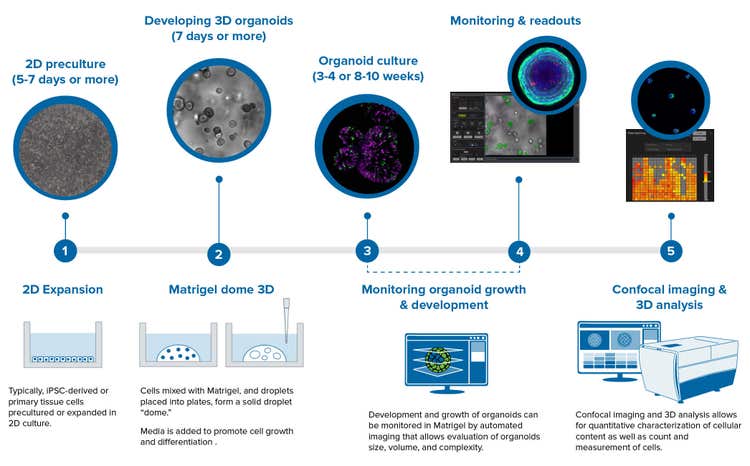
3D organoids can be derived from primary cells like intestinal organoids or induced pluripotent stem cells (iPSCs), e.g., neuronal or cardiac organoids. The workflow may start from 2D pre-culture or expansion of iPSC cells, followed by the differentiation step. After that, cells are mixed with Matrigel, and typically developed inside Matrigel domes, which also may include passaging and expansion. Intestinal organoids, colorectal, pancreatic, and liver are typically developed using this Matrigel dome step.

Alternatively, some other organoid types do not require Matrigel but instead are developed in low attachment plates (e.g., cardiac organoids).

Development of organoids takes from a few days to several weeks. Some protocols even require a few months. This is a very tedious process and will greatly benefit from automation.

Finally, the endpoint assay, whether it is a drug treatment, viral infectivity assay, or toxicity assessment, is set up in a multi-well format, with 96 or 384-well plates.

Next, cells are treated with drugs and processed for read-outs, which may include ATP assays, cell death assays, high-content imaging, or calcium oscillation.

4. Can you tell me a bit about how you apply 3D cell model workflows specifically to your research?

We are focusing on the development of automation protocols for automated cell cultures, as well as automated imaging and image analysis for complex 3D workflows. Recently, we developed and ran automated screening assays for finding more efficient anti-cancer drugs for triple-negative breast cancer. We used patient-derived cancer organoids representing a drug-resistant disease phenotype, and we applied automation to culture 3D organoids, simulate drug intervention, and run end-point assays for identifying the compounds that kill tumor cells. We tested a library of compounds and found several candidates that had greater efficacy than current standard drugs.

5. Can you explain how to automate the workflow for the development and analysis of organoids?

We created an automated workcell at Molecular Devices that combines several instruments in one complex system. It includes a Beckman Biomek automated liquid handler, a LiCONiC automated incubator, our ImageXpress HT.ai high-content imaging system, our SpectraMax plate reader and AquaMax washer, and a Bionex automated centrifuge. All the components are connected by a collaborative robot, PreciseFlex 400 that can move plates from one instrument to another at desired time points, while scheduling software ensures that all system elements work together seamlessly. Each instrument has multiple protocols designed for different steps, including feeding the cells and organoid plating, which can be called out by the scheduler at indicated times.

Organoid Innovation Center

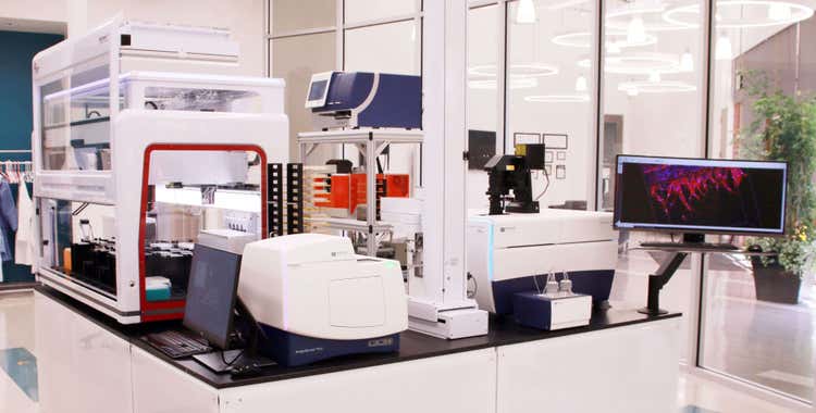
The Organoid Innovation Center at Molecular Devices combines cutting-edge technologies with novel 3D biology methods to address key challenges of scaling complex 3D biology.

Imaging methods are another exciting area of technology for organoid research. To image organoids or organ-on-a-chip, we need to use advanced optics. The ImageXpress high-content imaging system has several advantages for 3D samples:

Powerful lasers and confocal optics allow us to take the Z-stack of multiple images starting from the bottom and going up with steps like 5-10 microns Confocal optics allow us to reject the light that is out of focus so that we can get sharper images throughout organoids and Matrigel.

Next, our MetaXpress image analysis software analyzes the images in each 2D slice and converts data into 3D space. You can get multiple measurements to characterize organoids, cells, or subcellular organelles. These measurements help define cell counts, intensities, volumes, area, distances, and more, allowing us to monitor and quantitate changes in morphology, cell content, and viability. We also have machine learning elements, where users can train software to recognize objects, and features, to provide more efficient and insightful analysis.

6. How can automation aid in the research of complex systems specifically?

Automating would reduce labor and repetitive tasks like feeding cells every day or every second day for 2 months. Also, it will help to ramp up the research with higher throughput. For example, instead of studying 3 cell lines or 5 mutations, automation would allow you to test 50 cell lines to study 100 mutations.

High-content imaging powered by machine learning algorithms will allow to observe and characterize a variety of changes in organoids and cells, providing multiple readouts and yielding a valuable set of information about cell growth, differentiation, cell cycle, death, apoptosis, gene expression, or activation of signaling pathways.

7. How will you use these systems again in your future research?

In addition to cancer biology studies, we are actively developing other workflows, including but not limited to intestinal organoids, stem cell workflow, cardiac organoids, and more.

8. How will the automation of 3D organoid analysis evolve in the future?

We believe, as biology evolves and the complexity of assays increases, automation will be increasingly important for better understanding disease mechanisms, accelerating drug discovery, and eventually finding better ways to treat diseases.

By developing new and more advanced technologies and instruments, we believe we will further contribute to the progress of life sciences.

Understanding the basic principles behind 3D organoids - and the current bottlenecks - is crucial to the successful development and utilization of these advanced models for drug discovery. 


Listen to the full podcast

If you'd like to learn more, please enjoy the full podcast, “Complex assays with Ian Shoemaker, Beckman Coulter Life Sciences & Dr Oksana Sirenko, Molecular Device." 


Drug Target Review · Episode 14 - Ian Shoemaker, Beckman Coulter & Dr Oksana Sirenko, Molecular Devices

Here they discuss a comprehensive outlook on the science of organoid research – discover how organoid models are developed, used within our experts’ research, and how the automation of 3D organoid analysis will evolve in the future, plus much more!


  • Share on LinkedIn
  • Share on Facebook
  • Share on Twitter
  • Share on Youtube

Recent posts

View All Posts
Spacer

How 3D Cell Models Will Shape the Future of Drug Discovery

Target discovery and drug development rely heavily on 2D cell and animal…

A roundtable discussion
Spacer

A case study for assay-ready patient-derived organoids (PDOs) and high-throughput 3D imaging to advance drug discovery

Introduction – the problem.  The average cost of bringing a new drug to…

Read case study
Spacer

Celebrating the art of science: A D+I interview with Daniele Tortorella

At Molecular Devices, we cultivate a culture where associates around the…

Celebrate D+I
Spacer

Advanced technology for automated 3D biology workflows #SLASEurope2022

SLAS Europe 2022, hosted numerous sessions packed with the latest…

View SLAS EU activities
Spacer

[Podcast] Challenges of traditional cell line development and emerging technologies to help regulate monoclonality

Advancements in genetic engineering and synthetic biology have allowed…

Listen to CLD podcast
Compare /

Microplate Reader

  • SpectraMax i3x
  • SpectraMax iD3 / iD5
  • SpectraMax M 시리즈
  • SoftMax Pro 소프트웨어

세포 이미징

  • ImageXpress Confocal HT.ai
  • ImageXpress Pico
  • IN Carta 이미지 분석
    소프트웨어

클론 스크리닝

  • ClonePix 2 포유동물 콜로니 피커
  • QPix 미생물 콜로니 피커

FLIPR System

AXON PATCH-CLAMP

실험실 자동화

GxP 컴플라이언스

  • 뉴스
  • 이벤트
2023년 4월 2일

Focus on Microscopy (FOM)

2023년 5월 9일

Imaging User Meeting 2023

2023년 6월 26일

MPS World Summit

행사 더 보기

2023년 2월 22일

Silver Sponsor Molecular Devices at Society for Laboratory Automation and Screening 2023 International Conference and Exhibition

2023년 2월 13일

Molecular Devices inks deal with HUB Organoids to advance automated intestinal organoid screening technology

2023년 2월 7일

Molecular Devices partners with SEED Biosciences to exclusively offer DispenCell Single-Cell Dispenser, expanding leadership in cell line development

뉴스 더 보기
뉴스레터 로더
  • 2023년 3월  
  • 2023년 2월  
  • 2023년 1월  

글로벌 본사

  • 주소 아이콘

    Molecular Devices, LLC.
    3860 N First Street
    San Jose, CA 95134

  • 연락처

    Phone: +1 800-635-5577

    Phone: +86-400-821-3787

    Phone: +44-118-944-8000

    Phone: +44-118-944-8000

    전화: +82-2-3471-9531

    Teléfono: +44-118-944-8000

    Telefono: +44-118-944-8000

  • 사무실 영업 시간

    Mon-Fri 8:00 am - 5:00 pm (PST)

    周一至周五,早上 9 点至下午 5 点 30 分

    Montag bis Freitag 8 h à 17 h (GMT)

    lundi à vendredi 8 h à 17 h (GMT)

    월~금, 오전 9시~오후 5시(한국표준시)

    de lunes a viernes 8 h à 17 h (GMT)

    lunedi al venerdì 8 h à 17 h (GMT)

  • 제품 영업

    Sales +1 877-589-2214

    售前咨询 : 400-821-3787 转 1

    Verkauf 00800 665 32860

    Ventes 00800 665 32860

    판매 +82-2-3471-9531

    Ventas 00800 665 32860

    Saldi 00800 665 32860

  • 기술 지원

    Support +1 800 635 5577

    售后咨询 : 400-821-3787 转 2

    Unterstützung +44-118-944-8000

    Support +44-118-944-8000

    지원 +82-2-3471-9531

    Apoyo +44-118-944-8000

    Supporto +44-118-944-8000

  • 자료 창고

    SpectraNet 고객 포털

바닥글 탐색

  • 이용약관
  • 개인정보 보호정책
  • 데이터를 판매하거나 공유하지 마십시오.
  • 온라인 이용약관
  • 상표
  • 지원
  • 연락처
  • 사이트맵
  • Danaher Life Sciences
  • 채용

©2023 Molecular Devices, LLC. 모든 권리 보유.

©2023 Molecular Devices, LLC. 美谷分子仪器(上海)有限公司保留所有权利。
All Rights Reserved 沪ICP备05056171号-1

barcode