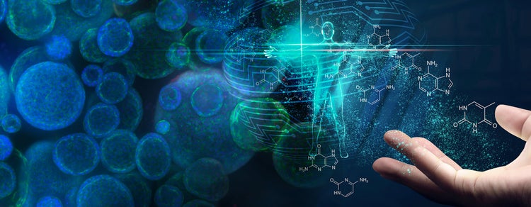
3D Cell Model로 신약 개발의 미래를 재편하는 방법
표적 개발과 신약 개발은 후보약물의 유효성 및 독성 효과를 판독하기 위해 2D 세포 및 동물 모델에 크게 의존합니다. Yet, 90% of candidates fail to make it past phase III clinical trials (1). This is often due to complications that preclinical models could not capture. Upgrading these models can circumvent the risk of clinical failure and prevent potential delays or termination of drug development projects. Replacing 2D cell models with self-organizing 3D models is one step towards this goal. However, this is not a simple process, and it introduces as many challenges as added benefits.
Drug Target Review discusses the past, present, and future of 3D cell models with Kenneth Pryde, Associate Director of Oncology Safety Sciences at AstraZeneca, Florian Fuchs, Chief Technology Officer at HeartBeat.bio, and Shan Dhamija, Vice President of Strategy and Innovation at Molecular Devices.
https://share.vidyard.com/watch/kfyFLgxVTWuQbgiTnZN2x4
In this roundtable discussion, leading experts from the industry discuss the promise of 3D biology and organoids for advancing drug discovery. They examine how to overcome challenges involved in 3D biology and what the future of organoid research will look like.
Are 2D cell cultures and animal models ideal during drug development?
Animal models are widely used for human biology studies and drug development. From a regulatory perspective and safety perspective, it is necessary for a drug molecule to undergo preclinical animal trials before phase 1 clinical trials. However, these preclinical observations have not always translated to clinical performance, due to limited assessment of pharmacokinetics and pharmacodynamics in animals as compared to humans. Dr. Pryde emphasizes the need for in vitro models: “Additional in vitro models are required to complement those animal models to help decrease the risk of attrition and safety issues manifesting in the clinic, which the animal models were not able to identify.”
Although 2D cellular models complement animal studies and improve preclinical assessment accuracy, their limitations surface with increasingly complex disease models and targeted drug discovery. Traditional 2D cell models and corresponding assays fail to recapitulate physiological events, such as cell-cell communication, cell-extracellular matrix (ECM) interactions, and physical constraints. According to Mr. Dhamija, a focus on new technologies is disproportionate: “While new tools allow precise and nuanced discovery at greater biological depth unless we change the model systems, we will only get better at finding the wrong answers.”
How do 3D cell models prevail over 2D cell models?
One of the main reasons 3D cell models stand out is that in contrast to 2D cell models, they can better represent the in vivo environment, making them more predictive of the potential effects of a compound. This also helps them perform better in toxicology studies, as they can preclinically predict potential risks. From this perspective, 3D cell models mitigate the risks associated with false negatives or positives in animal studies.
Since animal models are still indispensable in drug development and testing, 3D cell models can also be utilized to assess their relevance to human physiology. Cell models representing different animal tissues can help elucidate the correlation between animal and human cellular dynamics affecting immunogenic responses and functional alterations. The combination of types of models results in a more systemic view of the tissue or organ studied while minimizing the risk of data misinterpretation.
Different types of 3D cell models and their advantages
We can categorize 3D cell models into four main groups - spheroids, 3D bioprinted models, organoids, and organ-on-a-chip. Spheroids consist of a group of cells coalesced together to form spheres. 3D bioprinted models are a step above, as they introduce spatial complexity or extracellular matrices in addition to the cells. Organoids are tiny, self-organized three-dimensional tissue cultures that are derived from stem cells. The final category is organ-on-a-chip which implements microfluidics to simulate circulation, mechanical stressors, and the impact of interactions between different tissues.
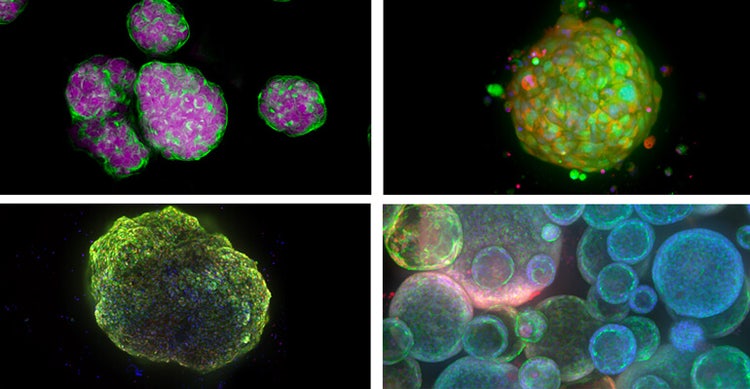
Examples of 3D cell models from the top left - Spheroids, patient-derived organoids (or tumoroids), cardioid (heart organoid), and lung organoids.
Each type of 3D model has its own advantages and disadvantages. However, organoids are widely used for a number of reasons. Dr. Fuchs favors organoids over other 3D models because “they have intrinsic capabilities for amplification, allowing the modeling of all developmental steps in a tissue”. However, he does acknowledge the limitations, such as the lack of an immunogenic component. Another highlight of organoids and spheroids, as emphasized by Dr. Pryde, is their scalability and high-throughput capability, as both are feasible for 384-well plates. This allows them to be regularly employed in high-throughput drug screening for efficacy and toxicology studies.
One of the main advantages of organoids is their versatility. One can tweak the organoid design to render a specific function through a huge repertoire of tools. Pluripotent stem cells are the primary source in organoid studies, as they can directly differentiate into tissue-specific cells. This makes it more biologically relevant and better at recapitulating the constituent cell types and extracellular matrix. Furthermore, the fact that these can be genetically altered enables the acquisition of deeper insights when studying disease mechanisms, drug action, drug resistance, and toxicology.
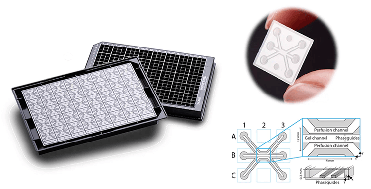
Mimeta’s organ-on-a-chip technology, OrganoPlate®
Future of organoid research
The emergence of biologically relevant organoids and technological refinements in organoid workflows create space for exciting research opportunities. Dr. Fuchs emphasizes the possible use of synthetic embryos to mimic embryo development, which could drive a better understanding of human development. He also addresses the need to transition to a completely animal-free preclinical testing routine, calling on regulatory parties to readjust their criteria. “In fact, animal models may not even be useful in testing biologics, such as vaccines for antibodies that are so humanized that they are weakly immunogenic in animal models. So, it does not make sense for us to use animals knowing that we will not be observing the impact of the therapeutic.”
Dr. Pryde anticipates that organoid research can immensely contribute to personalized medicine. “3D models create this new avenue in the therapeutic space around accessing biological material from patients to identify potential drug sensitivity or resistance. The results of this analysis can inform clinicians to decide whether a particular drug is beneficial and suitable for a patient or not”.
According to Mr. Dhamija, machine learning is likely to become a more integral part of 3D models, especially when derisking model systems. “While in academia, the incentive is for pursuing increased complexity, establishing robust 3D model research protocols is just as important for the industry. With machine learning, novel models can be made more reproducible, scalable, and consistent, which mitigates risk of data discrepancies that would cause significant delays in delivery to market and patients."
Nevertheless, organoid research has a long path to perfection. Of course, one of the challenges is gaining access to large patient samples or finding sufficient numbers of stem cell donors. Currently, drug screening and toxicology studies fail to translate to clinical studies because of patient variability. That’s why access to patient-derived material needs to be broadened to account for these variations in preclinical settings. Furthermore, to improve the credibility of predictions, researchers can complement in vitro models with computational models, which also brings a more holistic outlook to the disease of interest.
- Mullard, Asher. "Parsing clinical success rates." Nature Reviews Drug Discovery 15.7 (2016): 447-448.
* end *
Take a peek into where our research happens!
Tour our Organoid Innovation Center where we showcase cutting-edge instruments that work harmoniously together for autonomous, long-term, live cell 2D and 3D cell culture growth and monitoring with intelligent label-free imaging. This integrated workflow provides quality control alerts and readiness, 3D organoid screening, and deep-learning image analysis that reveals hidden patterns other technologies miss.