
Cell Counter
Cell Counter: Jurkat Cells
Jurkat 세포는 TCell Lymphocyte 백혈병에 걸린 남자 아이의 말초혈액으로부터 유래하였으며, 1970년대 후반에 확립된 immortalize T림프구 세포주입니다. 이 세포주는 T-cell leukemia, T-cell signaling, 그리고 Cancer Cell의 약물 민감도에 대한 연구를 위해 사용되어 왔습니다. 작고 둥근 세포로 Suspension에서 쉽게 생장하며, 여기에 제시된 것처럼 다양한 cell viability와 세포사멸 assay의 모델 시스템으로도 사용되었습니다.
StainFree Cell Detection 웨비나 보기
StainFree Cell Detection Application Note 다운로드
eBook 다운로드: 나도 Cell Counting 전문가
그림 1: StainFree 세포 계수
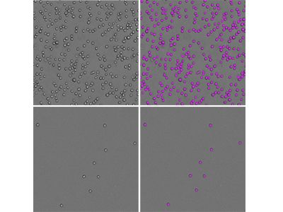
SpectraMax Minimax 300 Imaging Cytometer를 사용하여 Jurkat Cell을 이미징하였으며, 기본 설정 ‘CellsC’를 이용하여 세포를 식별하였습니다. 좌측에 보이는 것은 Transmitted Light에서 촬영한 원본 이미지이며, 우측은 같은 이미지를 소프트웨어로 식별하여 세포를 보라색 마스크로 나타낸 것입니다. 두 세포에 대한 밀도는 다음과 같습니다: 50,000개 및 1562개의 cell이 96 well plate에 well당 seeding되었습니다.
그림 2: StainFree 세포 계수 vs. 형광 세포 계수
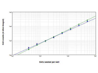
웰당 390~50,000개의 세포 밀도로 Jurkat 세포를 Seeding한 뒤 StainFree 기술(청색 점)을 이용하여 계수하거나 EarlyTox™ Live Cell Assay 시약을 사용하여 염색한 뒤 녹색 형광 세포를 계수하였습니다(녹색 점). 두 가지 방법으로 얻은 세포 수를 비교했을 때 Cell Density가 높은 일치도를 보입니다. 세포 계수는 웰당 4개 부분을 이미징하여 측정하였습니다.
그림 3: EarlyTox™ Live/Dead Assay
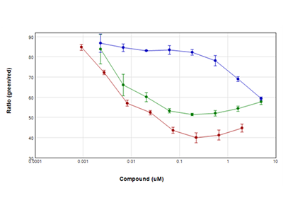
Jurkat Cell은 Staurosporine (적색 그래프), Camptothecin (녹색 그래프), Etoposide (청색 그래프)로 Serial Dilution 하여 24시간 동안 처리한 뒤, Molecular Devices의 EarlyTox Live/Dead Assay 키트를 이용해 assay를 수행하였습니다 . Live Cell은 Calcein AM(Green)로, Dead Cell은 ethidium homodimer(Red)로 염색하였으며, SpectraMax i3x Multi-Mode Microplate Reader의 Well Scan 기능을 이용하여 Well을 규칙적으로 가로질러 Multiple Read를 진행했습니다. 결과는 녹색/적색 비율과 처리된 약물의 농도를 비교하여 그래프로 나타냈습니다. 이 실험에 사용된 모든 약물 24시간 내에 cell viability를 크게 감소시켰습니다.
그림 4: EarlyTox Caspase-3/7 NucView 488 Assay
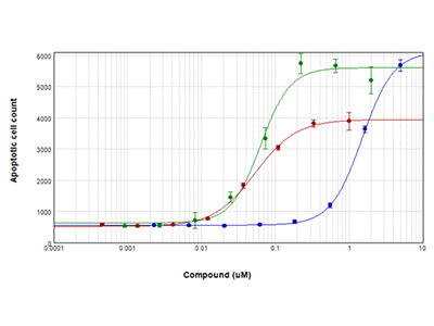
Jurkat 세포는 Staurosporine(적색 그래프), Camptothecin(녹색 그래프), Etoposide(청색 그래프)로 28시간 동안 처리한 뒤, EarlyTox Caspase-3/7 NucView 488 Assay 키트를 이용해 Apoptosis에 대한 분석을 시행했습니다. Caspase-3/7이 발현된 세포는 녹색 형광으로 염색하였으며, SpectraMax MiniMax 300 Imaging cytometer를 이용하여 이미징하고 계수했습니다.
Tip:
Jurkat 세포는 동그란 모양으로 균일한 형태를 보이기 때문에 이미지를 통해서 쉽게 계수할 수 있습니다. StainFree Cell Counting 에는, SoftMax Pro Software에서 기본 설정인 'CellsC'를 이용할 수 있습니다.
Jurkat Cell 분석에 필요한 장비와 시약
- SpectraMax ® i3x Multi-Mode Microplate Detection Platform
- SpectraMax ® MiniMax™ 300 Imaging Cytometer
- SoftMax ® Pro Software
Imaging Cytometer
Analysis type: Discrete Object Analysis
Wavelength for finding objects: TL
StainFree Cell Detection 기술에 관하여
Imaging을 기반으로 한 Cell-Based Assay는 일반적으로 형광물질을 세포에 붙여야 하는데, 이는 살아있는 세포에 독성이 될수도 있고, 혹은 Fixed Cell을 사용하여 염색을 해야 하기도 합니다. Cell Counting, Cell Confluence를 분석하는 경우, Label Free기법을 이용하면, Cell Viability에 영향을 주지 않고 Cell Proliferation과 Cell Viability를 정량적으로 Monitoring할 수 있습니다.
MiniMax 300 Imaging Cytometer가 장착된 SpectraMax i3 Multi-Mode Microplate 플랫폼에서 특허 출원 중인 유일무이한 기술, StainFree Cell Detection 기술을 이용할 수 있습니다. DNA에 삽입되는 DAPI나 장기적으로 세포에 독성이 되기도 하는 Live Cell Dye 등으로 세포를 염색하지 않고, 세포 생장, 세포독성 및 다른 assay를 수행할 수 있습니다.