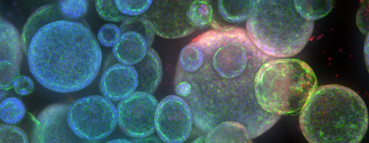
High-Throughput 3D Imaging의 도전과제를 극복
최근 Imaging 기술의 발전으로, 이제 3차원에서 복잡한 세포 네트워크를 관찰하고 분석할 수 있게 되었습니다. Through 3D imaging, we can acquire and analyze interactions in cells and tissues with greater detail and higher accuracy. 그러나 3D Imaging은 긴 이미지 스캐닝 시간부터 저해상도와 부적절한 분석툴에 이르는 여러 복잡한 문제가 있는 난해한 프로세스입니다.
Here is a summary of the common challenges faced in 3D cell imaging and how you can overcome them with the help of Molecular Devices products.
Out-of-focus light
Getting crisp 3D images can be challenging, especially if you are using widefield microscopy. Despite providing quick image acquisition, widefield microscopy falls short in eliminating out-of-focus signals. This leaves you with blurry images with intense out-of-focus light, hindering your image analysis.
The alternative is a confocal microscope with a pinhole that decreases sensitivity to out-of-focus light as you move away from the in-focus area, generating sharper images. The problem arises when a single pinhole needs to scan the entire image, which is time-consuming.
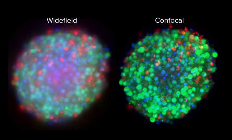
That’s where a spinning disk confocal microscope (SCDM) can serve as an upgrade. Instead of a single pinhole, an SDCM consists of hundreds of pinholes that rotate rapidly to scan the image, leading to a better resolution in a short amount of time. AgileOptix Spinning Disk Confocal Technology is a perfect example of this.
The ImageXpress® Micro Confocal High-Content Imaging System features the revolutionary AgileOptix™ Spinning Disk Confocal Technology that takes it one step further with a high-intensity laser light source for deeper tissue penetration, so you can obtain high-resolution images even from thick tissue samples. As a result, you can achieve a sharper image with improved cell visibility and at least a 30% increase in cell count.
Long acquisition time
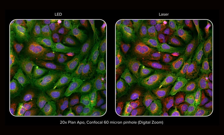
Before 3D laser scanning technologies, obtaining high-quality images required prolonged exposures, which was a challenge because of the 3D images drifting away from the center of the well. Today, confocal microscopes tackle this problem by implementing high-intensity laser sources.
High-intensity sources have several benefits that not only improve image quality but also increase the scan speed by reducing exposure time.
For example, the ImageXpress® Confocal HT.ai High-Content Imaging System boasts a seven-channel laser light source with eight imaging channels that can reduce the exposure time by up to 75%, which increases the scan speed by two-fold overall.
This especially comes in handy with yellow or cyan fluorescent protein imaging that requires the imaging of complex protein networks with intercellular and intracellular heterogeneities.
Reliable automated focusing
Maintaining focus on samples with thermal and mechanical fluctuations is challenging and can disrupt time-lapse imaging in particular. That’s why your microscope should automatically detect and stabilize focal planes. Digital camera-based autofocus systems are time-consuming because there is a prefocus process with an intricate search over a wide focusing range.
With recent advancements in laser autofocus technologies, you can accelerate automated focusing on various types of plates, including organ-on-a-chip and U-bottom.
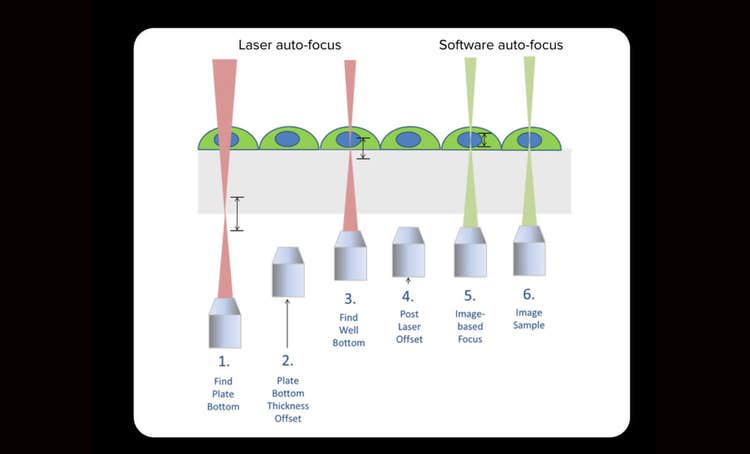
In particular, a hybrid autofocusing system comprising laser and image-based autofocus will generate the best results. This type of autofocus is much faster because the laser is flashed only once per well. Yet, you can still benefit from the sample compatibility of image-based focusing. Most importantly, the minimum amount of laser exposure means that you are minimizing the risk of phototoxicity, which is critical in live cell assays.
Analysis bottleneck
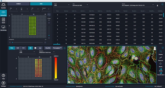
Data sets are often large and complex, so their analysis can take hours to complete. Overcoming this hurdle requires cutting-edge image analysis tools with accelerated processing, accurate image classification, and a simple user interface.
Molecular Devices image analysis tools stand out for their features that solve a variety of analysis needs.
For example, did you know you could speed up your time-lapse analysis by up to 40 times with multi-threaded parallel processing? This has become a reality with our MetaXpress® High-Content Image Acquisition and Analysis Software for both 2D and 3D imaging. Another challenge in image analysis is the classification of cell populations for comprehensive cell characterization. Here is where machine learning comes in. Our IN Carta™ Image Analysis Software enables automatic phenotyping classification in large and complex cellular profiles.
In both analysis tools, you can run processes with a variety of samples in a matter of minutes with a superb user experience.
Difficult light penetration
Perhaps one of the most difficult tasks in 3D imaging is tissue penetration, which is critical for obtaining information about the complex biological behavior of your sample. If your microscope has limited penetration depth, the image quality will decrease as the light gets scattered or absorbed in thick tissue samples.
As we mentioned earlier, confocal microscopy can provide better resolution, but further developments can translate this success to deep tissue penetration.
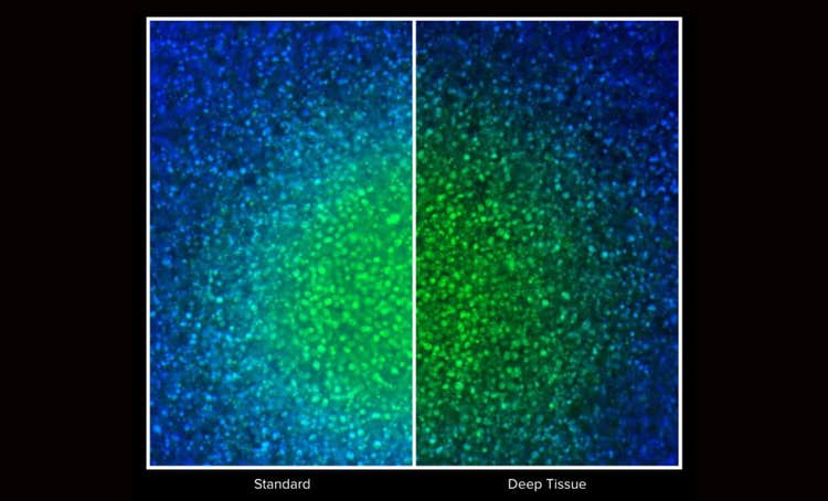
The microscope’s contrast ability is critical for the suppression of out-of-focus light, which allows better detection of the fluorescence emitted from the specimen. This can be achieved by a pinhole that rejects the out-of-focus signal. The contrast can be achieved even faster if you have multiple pinholes that are in constant communication with each other during image scanning.
Finally, combining confocal microscopy with high-intensity laser optics allows deeper penetration by sending longer wavelengths of light to excite fluorescence samples without damaging the sample or scattering the light.
High content data-mining
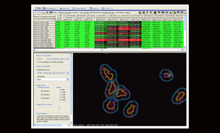
Imagine trying to visualize multi-well or multi-plate data. With a stand-alone 3D imaging software, you would probably have to switch between applications to view multiple image thumbnails and heatmaps.
To make data mining more practical, you need informatics software that can integrate image acquisition with image analysis and informatics so you can analyze data directly from the original image. With that purpose in mind, Molecular Devices developed AcuityXpress™ High-Content Informatics Software.
The software boasts an interactive zoom feature that allows you to navigate smoothly between images and numerical data. With this technology, you are in the driver’s seat when determining the level of detail, whether it is zooming into an individual high-quality image or an overall viewing of all images and data on one screen.
Besides the state-of-art navigation, AcuityXpress software provides a plethora of analysis and calculation tools for you to interpret complex data and multiple parameters at once.
Conclusion
3D imaging can bring deeper insights into the complex behavior of your samples; however, it comes with some challenges. At Molecular Devices, we strive to accompany you on every step of the imaging process to help guide you to your specific solution—from getting sharper images in minutes to organizing them in a centralized software system, we help you advance discovery.
Learn more about High-Content Screening with AgileOptix Technology and its combination of powerful solid-state lasers, water immersion objectives, scientific CMOS sensor, and dual spinning disk with five different disk geometries. Or contact us for more information.