어떻게 세포 염색이 신약 개발에서 중요하게 되었나
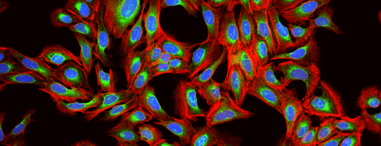
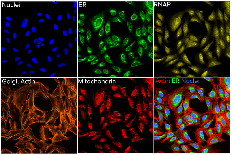
“백문이 불여일견”이라는 오래된 속담을 들어본 적이 있나요? When it comes to Cell Painting, this saying is especially true. 세포 염색은 세포학적 프로파일링에 사용되는 High-Content, 다중 이미지 기반 Assay입니다. The idea is to “paint” or stain as much of the cell as possible to get a representative image of the cell. Automated image analysis software is then used to extract hundreds to thousands of quantitative features of the cell, providing a rich phenotypic profile. Here, we shed some light on this emerging high-content image-based assay and the impact it is having on drug discovery.
Advancing the field of drug discovery
In recent years, Cell Painting has gained popularity across a wide range of research areas. For example, you can knock down or overexpress genes and look at their phenotypic profile to get a handle on the functions and the pathways of a specific gene.
Drug discovery is another area where Cell Painting is really making significant strides. It can be used to identify targets, toxicity in molecules, or compounds that are being studied. It can also be used to gain insights into different mechanisms of action or signaling pathways. In addition, you can test compounds in different cell lines and assess drug effects on potentially different organs. There are studies in which Cell Painting was used to screen pharmacological compounds in breast cancer cell lines. In these instances, the potential application is in the context of personalized medicine. By looking at the profiles that these compounds produce, researchers could identify the most suitable treatment for a specific clinical subtype.
As we all know, the drug discovery process is very costly. It typically takes about ten years and has a high attrition rate, making it very difficult to bring a drug to market. Recursion Pharma is a great example of a company that’s really leading the way in advancing the drug discovery process through Cell Painting. They implemented their own special machine learning pipeline to identify potential hits for drug discovery. The company was founded in 2013 and they already have four candidates in Phase 2 clinical trials and another six candidates in Phase 1.
Advantages of using Cell Painting over traditional cellular labeling assays
The Cell Painting assay is also highly adaptable to different applications, from studying gene function to identifying potential therapeutic compounds for SARS-CoV-2. It has also been demonstrated to be suitable for use on different cell types. This potentially is useful for assessing the effects of compounds or environmental chemicals on the human physiology.
In essence, the Cell Painting assay is very data rich, highly multiplexed and as thorough as it can be. The Carpenter lab at the Broad Institute summarizes it best, “Measure everything, ask questions later.”
* The standard Cell Painting assay referenced here is from Gustadottir et al 2013. Since then, there were other works published using different dyes suitable for their specific study.
Choosing the right high-content imaging system and analysis software for Cell Painting
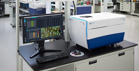
Our imagers are highly flexible and can be configured to meet specific assay requirements with widefield and various confocal options. One of the bottlenecks for Cell Painting is in imaging speed, because images are acquired from a 384-well plate and nine sites are typically imaged in each well. Multiply this by five channels and you get a huge amount of data. We offer solutions that help to improve image acquisition speeds, such as different cameras and laser light sources. Our instruments are also compatible with robotic automation which transfers plates in and out of the imager so that you can continuously generate data.
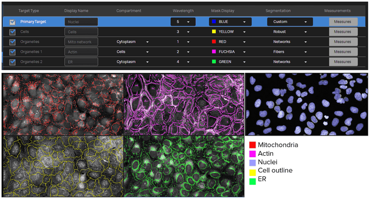
Feature extraction using IN Carta software analysis protocol
Because a Cell Painting assay produces objects of various shapes and sizes that are typically crowded or clumped together, unbiased feature extraction (i.e., segmentation) can be an obstacle for the image analysis portion of the workflow. To help overcome this challenge, our IN Carta™ Image Analysis Software includes a deep machine learning module called SINAP, which allows for more accurate image segmentation and greater reproducibility of data. And because the SINAP algorithm learns to segment by having users simply draw on the image, it is accessible to a wide range of users—from novices to more highly skilled software experts.
Learn more about our high-content imaging and analysis solutions that are used for Cell Painting. View ImageXpress Confocal HT.ai system and IN Carta software pages.