자동화된 오가노이드 세포 배양이 개발, Imaging, 분석되는 방법
In our latest post, scientists at Molecular Devices discuss advances in cell technologies and demonstrate an integrated workflow that allows automated processes of cell culture and imaging to monitor the development and characterize the complex responses in 3D organoids.
High-content imaging methods for automated cell culture organoids
When using organoids for disease modeling and assessment of compound effects, the quality of images is important for downstream analysis. For maximum quantitative and robust assessment of phenotypic changes in organoids, as well as for increasing throughput of experiments and screens, high-performance, automated imaging and analysis solutions are of critical importance.
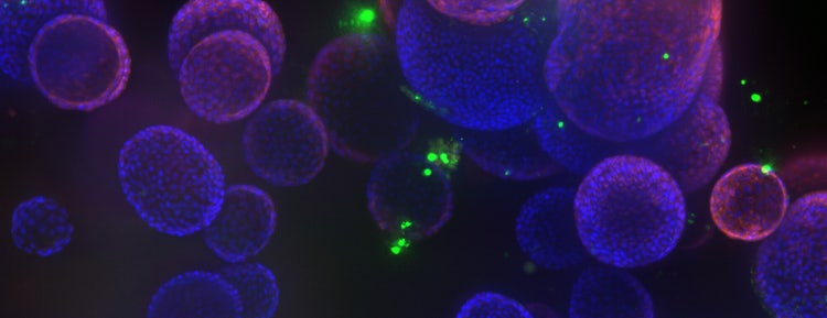
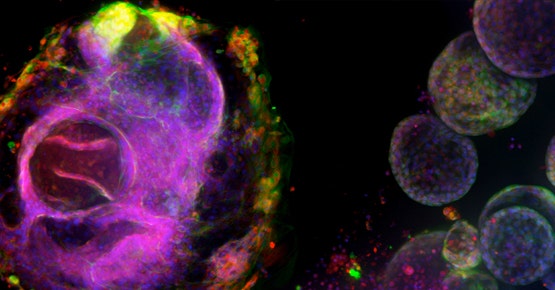
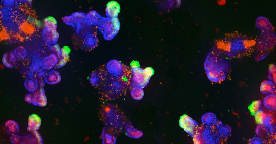
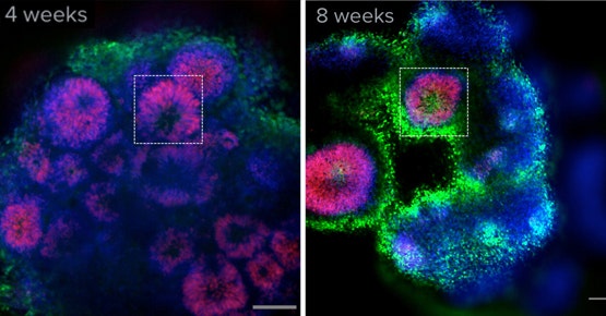
Confocal microscopy enables efficient imaging of 3D objects, including spheroids, organoids, and organ-on-a-chip models. A confocal microscope, such as the ImageXpress® Confocal HT.ai High-Content Imaging System utilizes a seven-channel laser light source with eight imaging channels to enable highly multiplexed assays; spinning disc confocal technology to penetrate deeper into thick tissue samples for sharper images; and water immersion objective increase signal to noise ratio, improve z-resolution, and decrease optical aberrations for sharper, crisper images
기존의 이미지 분석 방법은 수동 또는 반자동 방식으로 수행하게 되면 엄청나게 복잡하며 시간이 많이 소요될 수 있습니다. 작업이 복잡하며 그 세부적인 특성으로 인해 항상 사람이 실수하거나 비뚤림이 존재할 수 있습니다. When you add to this the repetitive, lengthy, and often laborious nature of the workflow, there comes the opportunity to apply advanced image analysis tools and AI/machine learning.
Advanced image analysis software would provide information about changes in phenotypes. MetaXpress® High-Content Image and Acquisition and Analysis Software allows users to find and characterize spheroids and then count and characterize cells inside spheroids, as well as sub-cellular objects. You can assess the specifics of 3D cell cultures, such as volume, diameter, shape, and intensity, which also helps you classify and organize data according to organoid features.
IN Carta® Image Analysis Software is a deep learning-based image segmentation tool enables robust label-free organoids and cell analysis. The machine learning tools can convert complex image data into actionable results. This solution helped researchers classify organoids based on size and diameter.
Overall, our proprietary AgileOptix technologies in confocal microscopy boast the qualities that can elaborately map out the complex structure of 3D organoids.
Organoid applications for drug discovery and development
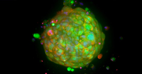
Organoids are becoming increasingly important in the fields of cancer research, neurobiology, stem cell research, and drug discovery, since they allow for the enhanced modeling of human tissues. Derived from stem cells, organoids can be differentiated into a wide range of tissue types including the lung, brain, and intestine to name a few. Because these 3D microtissues mimic in vivo organs, they can provide researchers with greater insight into the mechanisms of human development and disease, for example:
폐 오가노이드
Intestinal (gut) organoids
뇌(대뇌) 오가노이드
PDO/Tumoroids
Patient-derived organoids (PDOs) – or tumoroids – are 3D cultures that can be generated from primary tumors of individual patients. 튜머로이드는 암 연구, 신약 개발, 개인 맞춤의학에 있어 매우 가치 있는 도구입니다.
For example, efficient cancer therapy is crucial in the survival of cancer patients. This necessitates the use of clinically-relevant tumor models to understand the biology of disease, analyze tumor biomarkers, screen for the most efficient anti-cancer drugs, and provide a platform to study responses to targeted therapies.
Automation protocol of 3D biology workflows
Due to the complexity of organoids, more sophisticated 3D imaging and analysis techniques are needed to characterize these biological assays accurately and efficiently. 오늘날, 자동화 Confocal Imaging 시스템과 3D 이미지 분석 소프트웨어는 연구자들의 실험과정을 간소화하고 최적의 결과를 얻도록 돕기 위해 일반적으로 사용됩니다.
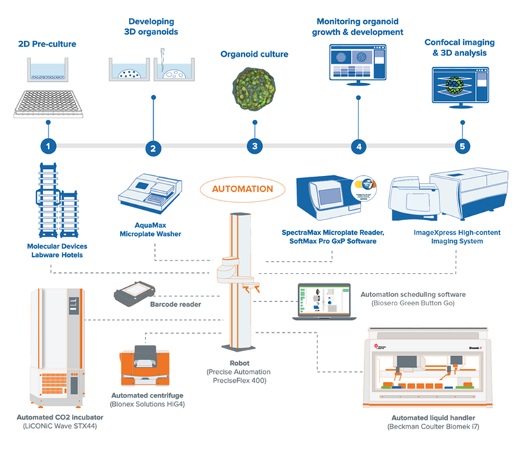
Our organoid screening workflow demonstrates an end-to-end method that utilizes leading technology from across the industry for automated cell culture, monitoring, and high-content imaging. The integrated workcell includes our SpectraMax® microplate reader, Aquamax microplate washer, ImageXpress® Confocal high-content imagers, automated CO2 incubator, automated liquid handler, as well as a collaborative robot. With intuitive scheduling software, researchers can control the 3D workflow for automating the seeding, media exchange, and monitoring of organoids development. In addition, the method allows for automation of compound testing and evaluation of phenotypic changes.
At our Organoid Innovation Center (OIC) we showcase these cutting-edge technologies with novel 3D biology methods to address key challenges of scaling complex 3D biology. 협동 스페이스에서 고객과 연구자들이 실험실에 참여하도록 하여 사내 과학자들의 지침과 함께 오가노이드 배양과 Screening을 위한 자동화 실험과정을 시험해 볼 수 있도록 합니다.