자동화된 3D 생물학 실험과정을 위한 고급 기술 #SLASEurope2022
SLAS Europe 2022, hosted numerous sessions packed with the latest research on emerging topics as well as sessions and panel discussions focused on how to build, and succeed, in a career within the life sciences or biotech industries.
We were delighted to attend SLAS EU 2022 in Dublin this year. We would like to thank all of you for visiting our booth, and for the opportunity to showcase our market-leading solutions to the scientific community. At the conference, we had the opportunity to deliver a series of educational presentations, together with our partners — From new advancements in engineering next-generation organoids to developing an automated lab workflow for 3D biology encompassing culturing, monitoring and high-content imaging.
Table of Contents
1. Automation of compound screening and high content imaging of 3D triple-negative breast cancer patient-derived tumoroids
Authors: Oksana Sirenko | Molecular Devices, LLC.
https://share.vidyard.com/watch/zbx5hvfDZWnGzvrWpoM9id
Finding efficient drug combinations to treat cancer patients is critical for therapy success, especially for rare types of tumors that resist traditional therapy. Triple negative breast cancer is a clinically aggressive subtype, with high rates of metastasis, recurrence, and drug resistance, and no clinically approved small molecule targeted therapies. There is a critical need to develop methods for efficient testing drug efficacy in patient-derived tumor samples to discover new therapeutic targets. Patient-derived 3D cancer models are highly valuable tools for cancer research and drug development. However, the complexity of performing 3D assays remains a hurdle for adopting these methods for compound screening. In the present study we describe automation of imaging, analysis, and cell culture methods which enables scaling up complex 3D cell-based assays and compound screening.
2. 생체모사칩 Assay의 자동화: 배양, Imaging, 혈관 형성 분석 자동화
Authors: Angeline Lim, Oksana Sirenko | Molecular Devices, LLC.
Arthur Stok, Matthew Delport | MIMETAS
Francis Enane | Beckman Coulter Life Sciences
https://share.vidyard.com/watch/yhhuxURXB5NdPd1jjC5s9J
There is a critical need for biological model systems that better resemble human biology in drug discovery. The OrganoPlate® was developed as an organ-on-a-chip platform allowing the formation of three-dimensional (3D), microfluidic-based, long-term cultures of live cells. However, the complexity of 3D models remains a hurdle for wide adoption in research and drug screening. Automation of the cell culture, assays, and analysis can provide the tools necessary to facilitate and scale up the use of organo-on chip systems.
3. 3D 인간 iPSC 유래 심장 삼중 배양 미세조직의 구조적 조직화와 화합물 반응의 기능적 분석
Authors: Simon Lydford1, Zhisong Tong1, Carole Crittenden1, Angeline Lim1, Sarah Himmerich2, Cara Rieger2, Ravi Vaidyanathan2, Coby Carlson2, and Oksana Sirenko1
Molecular Devices1
FUJIFILM Cellular Dynamics, Inc.2
https://share.vidyard.com/watch/uKsrsEjj6Gg72JCC3KzuAD
인간의 심장은 신체를 통해 혈액을 이동시키는 고도로 조절된 과정을 제공하는 복잡한 기관입니다. 성인의 심실은 심근세포, 내피세포, 섬유아세포, 기타 지지 세포 유형으로 구성됩니다. 심근세포는 인간 심실 전체 부피의 75%를 차지하지만 전체 세포 수의 50%만을 구성합니다. Recent publications show that tri-cellular co-culture microtissues of cardiomyocytes, endothelial cells and cardiac fibroblasts all derived from human iPSC enhance the maturation and functional activity of cells compared to 2D cardiomyocytes and thus more closely mimics actual heart physiology. In this study, we used a tri-culture model created by mixing iPSC-derived cardiac cells with primary adult fibroblasts and iPSC-derived endothelial cells at 70:20:10 ratio in ultra-low attachment (ULA) plates directly from thaw.
4. Assessing thrombin generation with the TECHNOTHROMBIN TGA kit on the SpectraMax i3x Multi-Mode Microplate Reader
Authors: Rebecca Kilmartin4, Cathleen Salomo3, Cathy Olsen2and Lieselotte Wagner1\ 1Technoclone Herstellung von Diagnostika und Arzneimitteln GmbH, 2Molecular Devices, LLC, 3Molecular Devices (Germany) GmbH and 4Molecular Devices (UK) Ltd
https://share.vidyard.com/watch/Qv2MqAxnDgV4cSrAE3Cbpe
Assessing the generation of thrombin in a plasma sample allows one to better understand coagulation mechanisms. To meet the needs of researchers to determine the time-dependent changes in thrombin concentrations on a flexible platform, Technoclone has developed a plate reader-compatible kinetic assay format, the TECHNOTHROMBIN® TGA kit, using a fluorogenic substrate. This poster demonstrates that the combination of the SpectraMax i3x reader and TECHNOTHROMBIN TGA kit offers an ideal platform to perform thrombin generation assays with good precision for research use.
5. Evaluation of a fluorescent approach to implement monoclonality assurance using calcein-AM viability dye.
Authors: Carola Mancini, PhD, Sarmad al-Bassam, PhD
Molecular Devices, LLC, Munich, Germany
https://share.vidyard.com/watch/cr8shNeTQEvYpKAUTfZUvv
Monoclonality 평가는 세포주 확립의 핵심이며 규제 기관은 생체의약품의 시장 출시를 위한 Monoclonality 근거를 요구합니다. Here, we demonstrate an optimized workflow using the fluorescence reagent, calcein-AM (CAM), in conjunction with a fluorescence-capable CloneSelect™ Imager (CSI-FL) that shows similar viability to label-free conditions while simultaneously providing high assurance of clonality. In this workflow, we determine a concentration of CAM that is ideal for detection of single cells on a CloneSelect™ Imager-FL while minimizing cytotoxic effects on clonal outgrowth. We describe guidelines for establishing an optimal concentration of viability dye for different cell types.
6. Novel 3D Tissue Models, Imaging and Automation of Organ-On-A-Chip Assays.
Authors: Oksana Sirenko - Molecular Devices, LLC,
Chiwan Chiang - MIMETAS
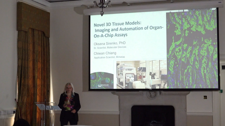
To enhance drug discovery & development, there is a critical need for complex biological model systems that better resemble human biology. In this tutorial, we provide an overview of complex 3D tissue models & assays, such as immune cell migration, and an introduction to high content imaging and image analysis methods, showing how you can get powerful new insights from complex biological models.
Explore solutions for automating your 3D biology workflows
Whether you’re just starting to explore the benefits of 3D tissue models and imaging or leveraging advanced 3D workflows, we offer solutions for the complexities that come with organoid acquisition and analysis.
Visit our Organoid Innovation Center to discover our newest collaborative space bringing customers, researchers, and in-house scientists together to test automated workflows for organoid culturing and screening. Automate your 3D culture workflows and quickly adopt innovative, 3D biological methods and technologies to reduce lab costs and push the boundaries of your research.