High-content analysis and phenotypic characterization of 2D and 3D cellular models
As presented by Kayla Hill, PhD, Field Applications Scientist, Molecular Devices
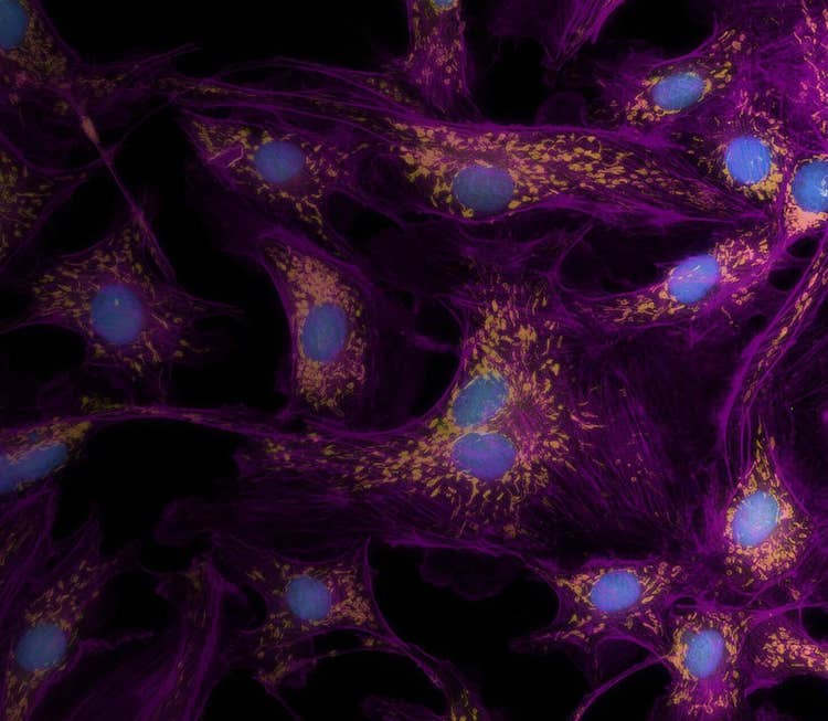
There is an increased need for expanding the variety and complexity of cell-based assays for biologic research and drug discovery. This has generated increasing interest in using three-dimensional (3D) cultures for assay development and phenotypic screening applicable for a range of cellular models including CNS, tumor micro-tissues, and organoids screens.
Combining these 2D and 3D models with live-cell assays allow monitoring of cell responses in real time and provide important insights about compound treatment effects, biological complexity, and physiological relevance of assay results. High-content imaging and analysis with the ImageXpress® Micro Confocal enables quantitative characterization of 2D and 3D cellular models. The state-of-the-art optics and powerful analysis tools generate phenotypic data from complex models in a high-throughput and automated manner.
Key highlights:
- Use high-content analysis to analyze multiple parameters across high-throughput compound screens
- Overcome challenges in automating 3D organoid imaging with robust and flexible autofocusing methods and targeted imaging workflows
- Characterize compound effects on tumor spheroid phenotypes and volumetric changes in 3D cellular content with 3D image analysis algorithms
For more information on the complete line of Molecular Devices High Content Imaging Systems, please click here.
The download is on the way to you. Check your email