
Cell Counter
Cell Counter: Rat Aortic Endothelial Cell
Rat Aortic Endothelial Cell은 Cell-cell Adhesion, Migration, Angiogenesis뿐 아니라 정상세포와 질병이 있는 세포의 Endothelial Function에 관련된 Cellular Signaling Pathway 등 다양한 과학적 연구를 위해 사용되어 왔습니다. 다만 이 세포는 Primary Cell 이므로 신약 후보물질 스크리닝에 사용하기는 어려울 수 있습니다. Assay를 성공적으로 수행하기 위해서는 assay 전 live cell imaging과 Cell Confluence를 측정하는 것이 중요합니다.
StainFree Cell Detection 웨비나 보기
StainFree Cell Detection Application Note 다운로드
eBook 다운로드: 나도 Cell Counting 전문가
그림 1. Rat Aortic Endothelial Cell의 Transmitted light Image
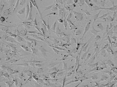
Single Primary Culture에서 Cell Process와 Cell Morphology를 보여줍니다.
그림 2. Rat Aortic Endothelial Cell에서의 StainFree counting
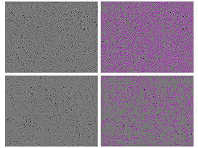
SpectraMax i3 Minimax 300 Imaging Cytometer를 사용하여 Rat Aortic Endothelial Cell을 이미징하였으며, Custom Analysis을 이용하여 세포를 확인하였습니다. 좌측 이미지는 Transmitted Light에서 촬영한 사진 원본(상단: 고밀도 배양, 하단: 저밀도 배양)이며, 오른쪽은 동일한 이미지에서 소프트웨어가 식별한 세포의 영역을 보라색 마스크로 나타낸 것입니다.
Image는 콜롬비아의 미주리 대학교 Dr. Virginia Huxley와 우리의 Field Application Scientist인 Randy Milano이 제공하였습니다.
Tip:
Rat Aortic Endothelial Cell은 매우 편평하고, 서로 다른 형태가 다중으로 나타나는 데다가, 때로는 그림 1과 같이 신경세포 증식물을 연상시키도록 길게 자라나는 모양으로 인해 이미징 기반의 세포분석이 어려울 수 있습니다. StainFree Technology는 여기에 Machine Learning 알고리즘을 적용하여 편의성을 높였습니다. 이미지 분석 설정에서 '새 설정 만들기' 옵션을 선택하고 세포 중심에 초점을 맞추어 그리기 기능을 사용하면, 세포 돌기(증식물)를 무시할 수 있습니다. 대표로 서로 다른 형태를 가진 일부 세포의 모양이 포함되도록 하면, 그림 2과 같은 결과를 얻을 수 있습니다.
Helpful hint: 초점을 맞출 때, Negative Value 쪽으로 더 조정하게 될 경우, 보다 뚜렷한 세포 이미지를 얻을 수 있어 소프트웨어를 이용한 Counting이 더 쉬워집니다.
Rat Aortic Endothelial Cell 분석에 필요한 장비와 시약
- SpectraMax ® i3 Multi-Mode Microplate Detection Platform
- SpectraMax ® MiniMax™ 300 Imaging Cytometer
- SoftMax ® Pro Software
장비 설정 보기
Imaging Cytometer
Exposure: 7ms
Focus adjustment: -455µm(사용된 세포의 특성에 따라 최적화될 수 있음)
Analysis type: Discrete Object Analysis
Wavelength for finding objects: TL
StainFree Cell Detection 기술에 관하여
Imaging을 기반으로 한 Cell-Based Assay는 일반적으로 형광물질을 세포에 붙여야 하는데, 이는 살아있는 세포에 독성이 될수도 있고, 혹은 Fixed Cell을 사용하여 염색을 해야 하기도 합니다. Cell Counting, Cell Confluence를 분석하는 경우, Label Free기법을 이용하면, Cell Viability에 영향을 주지 않고 Cell Proliferation과 Cell Viability를 정량적으로 Monitoring할 수 있습니다.
MiniMax 300 Imaging Cytometer가 장착된 SpectraMax i3 Multi-Mode Microplate 플랫폼에서 특허 출원 중인 유일무이한 기술, StainFree Cell Detection 기술을 이용할 수 있습니다. DNA에 삽입되는 DAPI나 장기적으로 세포에 독성이 되기도 하는 Live Cell Dye 등으로 세포를 염색하지 않고, 세포 생장, 세포독성 및 다른 assay를 수행할 수 있습니다.